. 2013 Dec 20;8(12):e84324.
doi: 10.1371/journal.pone.0084324. eCollection 2013.

Identification of a population of epidermal squamous cell carcinoma cells with enhanced potential for tumor formation

Affiliations

Identification of a population of epidermal squamous cell carcinoma cells with enhanced potential for tumor formation

Gautam Adhikary et al. PLoS One. .

Abstract

Epidermal squamous cell carcinoma is among the most common cancers in humans. These tumors are comprised of phenotypically diverse populations of cells that display varying potential for proliferation and differentiation. An important goal is identifying cells from this population that drive tumor formation. To enrich for tumor-forming cells, cancer cells were grown as spheroids in non-attached conditions. We show that spheroid-selected cells form faster growing and larger tumors in immune-compromised mice as compared to non-selected cells. Moreover, spheroid-selected cells gave rise to tumors following injection of as few as one hundred cells, suggesting these cells have enhanced tumor-forming potential. Cells isolated from spheroid-selected tumors retain an enhanced ability to grow as spheroids when grown in non-attached culture conditions. Thus, these tumor-forming cells retain their phenotype following in vivo passage as tumors. Detailed analysis reveals that spheroid-selected cultures are highly enriched for expression of epidermal stem cell and embryonic stem cell markers, including aldehyde dehydrogenase 1, keratin 15, CD200, keratin 19, Oct4, Bmi-1, Ezh2 and trimethylated histone H3. These studies indicate that a subpopulation of cells that possess stem cell-like properties and express stem cell markers can be derived from human epidermal cancer cells and that these cells display enhanced ability to drive tumor formation.


Conflict of interest statement

Competing Interests: The co-author, Richard L. Eckert, is a PLOS ONE Editorial Board Member. This does not alter the authors' adherence to all the PLOS ONE policies on sharing data and materials. Some of the authors on this manuscript, Shayne Boucher and Mohan Vemuri, are employed by Life Technologies. This does not alter the authors' adherence to all the PLOS ONE policies on sharing data and materials.

Figures

Figure 1
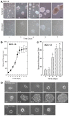
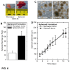
Figure 1. A subpopulation of SCC-13 cells grow as spheroids.
A SCC-13 monolayer cultures, maintained in growth medium, were harvested and plated at 40,000 cells per 9.5 cm2 in poly-HEMA coated dishes or in standard tissue culture plates and grown in spheroid medium. Monolayer and P1 spheroid cultures were monitored for growth. The bars = 50 μm. B SCC-13 spheroid growth rate. SCC-13 cells were plated on poly-HEMA coated dishes and the diameter of P1 spheroids was monitored for 0 - 9 d. The values are mean + SEM, n = 3. C Spheroids are formed from a subset of SCC-13 cells. SCC-13 cells (40,000 single cells) were plated on poly-HEMA coated dishes and the total number of P1 spheroids was monitored for 0 - 10 d. Care was taken to assure that spheroid formation was not due to cell aggregation. A small percentage of cells (0.15%) are able to form spheroids. The values are mean + SEM, n = 4. The asterisks indicate a significant increase in spheroid number as compared to the day 2 data point (p < 0.05). D Selection of spheroid-forming cells. SCC-13 monolayer cultures were dissociated and plated into 96 well non-attachment plates at one cell per well. At the indicated times after plating, the cells were photographed. The upper panel shows on of the 0.15% of cells that survived and formed a spheroid. The bottom panel shows one of the non-surviving cells undergoing cell death.
Figure 2
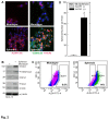
Figure 2. SCC-13 spheroid cultures express stem cell-related marker proteins.
SCC-13 cells, maintained in growth medium, were harvested and grown as P1 spheroids on non-attached (spheroid) or attached (monolayer) conditions. A Spheroid cultures express stem cell marker proteins. After 10 d, P1 spheroids and monolayer cultures were harvested, and the cells were permitted to attach on glass coverslips and stained with anti-ALDH1, anti-CD200 or anti-K5. No signal was observed in absence of primary antibody (not shown). B Spheroid cultures express stem cell marker proteins. Ten day spheroid and monolayer cultures were harvested for preparation of total extract, and the indicated epitopes were detected by immunoblot. C P1 spheroids are enriched in ALDH+ cells. P1 monolayer and spheroid cultures were grown for 10 d, harvested as single cell suspensions, incubated with ALDH1 substrate, and ALDH1 positive and negative cells were quantified by flow cytometry. The boxes indicate the sorting windows for cells considered ALDH+ and ALDH-. D ALDH1+ cells form spheroids. ALDH1(+) and ALDH1(-) cells, isolated from 10 d P1 spheroid cultures by cell sorting, were plated as a single cell suspension at 40,000 cells per 9.5 cm2 well on non-attachment dishes for a 10 d spheroid formation assay. At 10 d the number of spheroids were counted. The values are mean + SEM, n = 3. The asterisk indicates that spheroid formation by ALDH1(+) cells is significant greater as compared to ALDH1(-) cells (p < 0.05).
Figure 3
Figure 3. Stem cell marker expression in SCC-13 α6-integrin+/CD71- cells.
A Isolation of α6-integrin+/CD71- cells. Dissociated cells from 10 d spheroids were incubated with anti-α6 integrin beads to select for anti-α6 integrin positive cells which were then incubated with anti-CD71 beads to remove CD71+ cells to yield the α6-integrin+/CD71- cells. B Spheroid-derived α6-integrin+/CD71- cells express epidermis-relevant stem cell markers. α6-integrin+/CD71- cells were isolated using anti-α6-integrin and anti-CD71 beads as outlined above. These cells were then stained with anti-CD200 (green), anti-K15 (red), anti-K19 (green), anti-Sox2 (red), anti-Oct4 (green) and anti-ALDH1 (red) and visualized by confocal microscopy. Similar results were observed in each of three experiments.
Figure 4
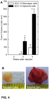
Figure 4. SCC-13 spheroid-selected cells display enhanced tumor formation.
A SCC-13 spheroid- or monolayer-derived cells were injected at 100,000 cells per site in NSG mice and tumor growth was monitored. Tumors volume was calculated as 4/3π x (diameter/2)3 [64]. The values are mean + SEM for 8 individual tumors. Asterisks indicate significant differences in tumor size between the spheroid and the monolayer groups at each time point (p < 0.005, n = 8). B Representative spheroid- and monolayer-derived tumors were harvested on week four and photographed. The spheroid-derived tumors are larger and red (vascularized) in appearance.
Figure 5
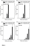
Figure 5. Spheroid-selected cells form tumors at lower injection densities.
SCC-13 spheroid- and monolayer-derived cells were injected at 100 to 100,000 cells per each of two or four sites in NSG mice (three to ten mice injected per cell number). Tumor formation was monitored by palpation and tumor size values are in cubic millimeters. The values are mean + SEM. Asterisks indicate significant differences in tumor size between the spheroid group and the monolayer group at each time point (p < 0.005).
Figure 6
Figure 6. Spheroid-selected skin tumor cells retain properties during in vivo growth.
A Morphology of tumors derived following injection of 100,000 spheroid-selected and non-selected (monolayer) cells after growth for four weeks. B/C Cells derived from spheroid-selected and non-selected tumors were dissociated and ability to form spheroids in culture was monitored. Spheroid formation is significantly greater for tumors formed from spheroid-selected cells. The values are mean + SEM, n = 4 independent tumors per group (p < 0.005). The bars = 100 μm. The photographs were taken after 10 d of spheroid growth. D Spheroid growth rate is identical for tumor cells derived from spheroid-selected and monolayer cells. Forty thousand cells, harvested from the tumors derived from spheroid-selected and non-selected cells, were plated in spheroid selection medium to monitor the rate of spheroid growth. Values are the mean + SEM (n = 4 independent tumors per group). No significant difference is observed in spheroid growth rate for cells derived from monolayer and spheroid tumors.
Figure 7
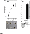
Figure 7. A431 skin cancer cells contain a population of stem cell marker-positive cells with enhanced ability to form tumors.
A A431 cells form spheroids. A431 cells were plated at 40,000 cells per 9.5 cm2 well in spheroid-selection medium and the rate of spheroid formation and morphology were recorded. The bottom panel is an image of P1 spheroids after 10 d of growth. B A subpopulation of A431 cells form spheroids. A431 cells (40,000) were plated in spheroid growth conditions and spheroid number was monitored on days 1 and 10. Among the 40,000 cells plated in this assay, only 0.03% survive and form spheroids. The asterisk indicates a statistically significant increase in spheroid number at day 10 compared to day 1 (p < 0.005, n = 3) D Spheroid-selected A431 cells express stem cell markers. Ten day spheroid and monolayer A431 cultures were harvested and extracts were assayed for expression of the indicated stem cell markers by immunoblot.
Figure 8
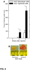
Figure 8. Spheroid-selected A431 cells display enhanced tumor formation.
A Spheroid- or monolayer-derived A431 cells were injected at 500,000 cells per site into each of the four sites in NSG mice and tumor growth was monitored. Tumor volume was calculated as 4/3π x (diameter/2)3 [64]. The values are mean + SEM for six individual tumors. Asterisks indicate significant differences in tumor size between the spheroid and the monolayer groups at each time point (p < 0.005, n = 8). B Spheroid- and monolayer-derived A431 cell tumors were harvested on week four and photographed. The spheroid-derived tumors are larger and appear vascularized (red).

Similar articles

Cited by

References

    1. Gupta S, Mukhtar H (2002) Chemoprevention of skin cancer: current status and future prospects. Cancer Metastasis Rev 21: 363-380. doi:10.1023/A:1021275330385. PubMed: 12549772. - DOI - PubMed
    1. Pincelli C, Marconi A (2010) Keratinocyte stem cells: friends and foes. J Cell Physiol 225: 310-315. doi:10.1002/jcp.22275. PubMed: 20568222. - DOI - PubMed
    1. Al-Hajj M, Clarke MF (2004) Self-renewal and solid tumor stem cells. Oncogene 23: 7274-7282. doi:10.1038/sj.onc.1207947. PubMed: 15378087. - DOI - PubMed
    1. Al-Hajj M, Wicha MS, ito-Hernandez A, Morrison SJ, Clarke MF (2003) Prospective identification of tumorigenic breast cancer cells. Proc Natl Acad Sci U S A 100: 3983-3988 - PMC - PubMed
    1. Singh SK, Hawkins C, Clarke ID, Squire JA, Bayani J et al. (2004) Identification of human brain tumour initiating cells. Nature 432: 396-401. doi:10.1038/nature03128. PubMed: 15549107. - DOI - PubMed

Publication types

Substances