. 2013 Dec 20;8(12):e84425.
doi: 10.1371/journal.pone.0084425. eCollection 2013.

Differentially expressed wound healing-related microRNAs in the human diabetic cornea

Affiliations

Differentially expressed wound healing-related microRNAs in the human diabetic cornea

Vincent A Funari et al. PLoS One. .

Abstract

MicroRNAs are powerful gene expression regulators, but their corneal repertoire and potential changes in corneal diseases remain unknown. Our purpose was to identify miRNAs altered in the human diabetic cornea by microarray analysis, and to examine their effects on wound healing in cultured telomerase-immortalized human corneal epithelial cells (HCEC) in vitro. Total RNA was extracted from age-matched human autopsy normal (n=6) and diabetic (n=6) central corneas, Flash Tag end-labeled, and hybridized to Affymetrix® GeneChip® miRNA Arrays. Select miRNAs associated with diabetic cornea were validated by quantitative RT-PCR (Q-PCR) and by in situ hybridization (ISH) in independent samples. HCEC were transfected with human pre-miR™miRNA precursors (h-miR) or their inhibitors (antagomirs) using Lipofectamine 2000. Confluent transfected cultures were scratch-wounded with P200 pipette tip. Wound closure was monitored by digital photography. Expression of signaling proteins was detected by immunostaining and Western blot. Using microarrays, 29 miRNAs were identified as differentially expressed in diabetic samples. Two miRNA candidates showing the highest fold increased in expression in the diabetic cornea were confirmed by Q-PCR and further characterized. HCEC transfection with h-miR-146a or h-miR-424 significantly retarded wound closure, but their respective antagomirs significantly enhanced wound healing vs. controls. Cells treated with h-miR-146a or h-miR-424 had decreased p-p38 and p-EGFR staining, but these increased over control levels close to the wound edge upon antagomir treatment. In conclusion, several miRNAs with increased expression in human diabetic central corneas were found. Two such miRNAs inhibited cultured corneal epithelial cell wound healing. Dysregulation of miRNA expression in human diabetic cornea may be an important mediator of abnormal wound healing.


Conflict of interest statement

Competing Interests: Co-author Dr. Alexander V. Ljubimov is a PLOS ONE Editorial Board member. This does not alter the authors' adherence to all the PLOS ONE policies on sharing data and materials.

Figures

Figure 1
Figure 1. Q-PCR validation of differentially expressed miRNAs.
Independent validation of differentially expressed miRNAs in ex vivo diabetic corneas by Q-PCR. N, normal; DM, diabetic. Note increased expression of three miRNAs in diabetic samples. Bars represent SEM.
Figure 2
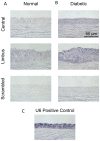
Figure 2. MiR-146a expression in the cornea.
In situ hybridization with locked nucleic acid (LNA) modified probes was performed on sections of normal (A) and diabetic (B) corneas compared to scrambled sequences (with Cy3-labeled pre-miRNA). Diabetic corneas show somewhat elevated expression both in the central cornea and limbus. In the diabetic corneas, the miR-146a signal is stronger in the limbus than in the central cornea. Positive control in normal cornea (with U6 miRNA) is presented in panel C. Bar = 60 μm.
Figure 3
Figure 3. Effect of miRNAs on wound healing.
A. HCEC were transfected with pre-miR-146a, -424, and -145 (top panel) or their antagomirs, pre-miRNA inhibitors (bottom panel); for control, irrelevant Cy3-labeled pre-miRNA was used. HCEC were scratch wounded as indicated in Methods. Wound closure was quantified using ImageJ software at 20 hr after wounding for pre-miRNA transfected cells and at 24 hr for pre-miRNA antagomir transfected cells. Bars represent SEM. *, p < 0.05 vs. miR-Cy3. B. miR-146a decreased wound closure, whereas its antagomir significantly increased it at 20 h compared to control. Values are expressed as percentage of the initial wound area. Data are means of three independent experiments in triplicate.
Figure 4
Figure 4. Western blot analysis of EGFR and p-EGFR expression in transfected HCEC.
Total extracted protein from HCEC transfected with pre-miRNA precursors or their inhibitors (antagomirs, AM) and Cy3-labeled pre-miRNA (control) was separated on gradient SDS-PAGE gels, transferred to nitrocellulose and probed with antibodies to EGFR or p-EGFR. Antibodies to β-tubulin or β-actin were used as equal loading controls and for semi-quantitation. In both wounded and non-wounded HCEC, miR-146a mimic treatment decreased whereas its antagomir increased protein levels of p-EGFR (A,B). MiR-424 mimic and antagomir treatments did not change the expression of p-EGFR in non-wounded cells (A,B). However, in wounded cells, miR-424 mimic decreased whereas its antagomir increased protein levels of p-EGFR (A,B). In both wounded and non-wounded HCEC, miR-146a mimic treatment decreased whereas its antagomir significantly increased protein levels of total EGFR (C,D). MiR-424 mimic and antagomir treatments did not significantly change the expression of EGFR both in wounded and non-wounded cells (C,D). Band intensities were quantified using ImageJ software and plotted relative to the loading controls. Blots in A were developed with alkaline phosphatase system, blots in C, with ECL reagent. *, p < 0.05.
Figure 5
Figure 5. Western blot analysis of p38 and p-p38 expression in transfected HCEC.
Total extracted protein from HCEC transfected with pre-miRNA precursors or their inhibitors (antagomirs, AM) and Cy3-labeled pre-miRNA (control) was separated on SDS-PAGE gels, transferred to nitrocellulose and probed with antibodies to p38 or p-p38. Antibodies to β-actin or β-tubulin were used as equal loading control and for semi-quantitation. In both wounded and non-wounded HCEC, miR-146a mimic treatment decreased whereas its antagomir increased protein levels of p-p38 (A,B). MiR-424 mimic and antagomir treatments did not change the expression of p-p38 in non-wounded cells (A,B). However, in wounded cells, miR-424 mimic decreased whereas its antagomir increased protein level of p-p38 (A,B). MiR-146a and miR-424 mimic and their antagomir treatments did not change the expression of total p38 both in non-wounded (C) and wounded cells (D). Band intensities were quantified using ImageJ software and plotted relative to the loading controls. Vertical lines in C and D separate different parts of the same gel. Blots were developed with alkaline phosphatase system. *, p < 0.05.
Figure 6
Figure 6. MiR-424 and wound healing-related signaling molecules.
In wounded HCEC cultures, miR-424 elevated in diabetes decreased staining for p-p38, p-Akt and especially, for p-EGFR. Conversely, antagomir (AM) treatment increased the expression of all tested activated signaling intermediates above control levels. Exposure times are the same for all panels in a row. W, wound area. Bar = 60 μm.

References

    1. Hamilton AJ, Baulcombe DC (1999) Species of small antisense RNA in posttranscriptional gene silencing in plants. Science 286: 950–952. doi:10.1126/science.286.5441.950. PubMed: 10542148. - DOI - PubMed
    1. Aravin AA, Lagos-Quintana M, Yalcin A, Zavolan M, Marks D et al. (2003) The small RNA profile during Drosophila melanogaster development. Dev Cell 5: 337–350. doi:10.1016/S1534-5807(03)00228-4. PubMed: 12919683. - DOI - PubMed
    1. Lund E, Güttinger S, Calado A, Dahlberg JE, Kutay U (2004) Nuclear export of microRNA precursors. Science 303: 95-98. doi:10.1126/science.1090599. PubMed: 14631048. - DOI - PubMed
    1. Maroney PA, Yu Y, Fisher J, Nilsen TW (2006) Evidence that microRNAs are associated with translating messenger RNAs in human cells. Nat Struct Mol Biol 13: 1102–1107. doi:10.1038/nsmb1174. PubMed: 17128271. - DOI - PubMed
    1. Brennecke J, Hipfner DR, Stark A, Russell RB, Cohen SM (2003) Bantam encodes a developmentally regulated microRNA that controls cell proliferation and regulates the proapoptotic gene hid in Drosophila. Cell 113: 25-36. doi:10.1016/S0092-8674(03)00231-9. PubMed: 12679032. - DOI - PubMed

Publication types