. 2013 Dec 23;8(12):e84433.
doi: 10.1371/journal.pone.0084433. eCollection 2013.

Defining the role of fluid shear stress in the expression of early signaling markers for calcific aortic valve disease

Affiliations

Defining the role of fluid shear stress in the expression of early signaling markers for calcific aortic valve disease

Ling Sun et al. PLoS One. .

Abstract

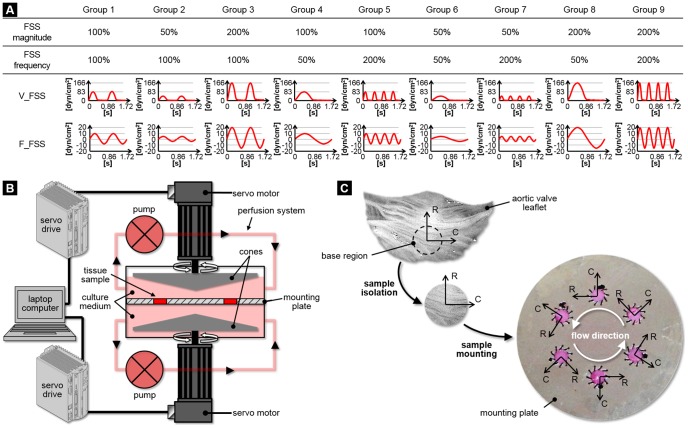
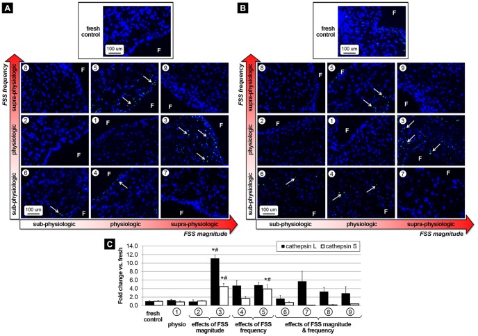
Calcific aortic valve disease (CAVD) is an active process presumably triggered by interplays between cardiovascular risk factors, molecular signaling networks and hemodynamic cues. While earlier studies demonstrated that alterations in fluid shear stress (FSS) on the fibrosa could trigger inflammation, the mechanisms of CAVD pathogenesis secondary to side-specific FSS abnormalities are poorly understood. This knowledge could be critical to the elucidation of key CAVD risk factors such as congenital valve defects, aging and hypertension, which are known to generate FSS disturbances. The objective of this study was to characterize ex vivo the contribution of isolated and combined abnormalities in FSS magnitude and frequency to early valvular pathogenesis. The ventricularis and fibrosa of porcine aortic valve leaflets were exposed simultaneously to different combinations of sub-physiologic/physiologic/supra-physiologic levels of FSS magnitude and frequency for 24, 48 and 72 hours in a double cone-and-plate device. Endothelial activation and paracrine signaling were investigated by measuring cell-adhesion molecule (ICAM-1, VCAM-1) and cytokine (BMP-4, TGF-β1) expressions, respectively. Extracellular matrix (ECM) degradation was characterized by measuring the expression and activity of the proteases MMP-2, MMP-9, cathepsin L and cathepsin S. The effect of the FSS treatment yielding the most significant pathological response was examined over a 72-hour period to characterize the time-dependence of FSS mechano-transduction. While cytokine expression was stimulated under elevated FSS magnitude at normal frequency, ECM degradation was stimulated under both elevated FSS magnitude at normal frequency and physiologic FSS magnitude at abnormal frequency. In contrast, combined FSS magnitude and frequency abnormalities essentially maintained valvular homeostasis. The pathological response under supra-physiologic FSS magnitude peaked at 48 hours but was then maintained until the 72-hour time point. This study confirms the sensitivity of valve leaflets to both FSS magnitude and frequency and suggests the ability of supra-physiologic FSS levels or abnormal FSS frequencies to initiate CAVD mechanisms.


Conflict of interest statement

Competing Interests: The authors have declared that no competing interests exist.

Figures

Figure 1
Figure 1. Experimental methods.
Side-specific FSS conditions considered in this study (A), schematic of the shear stress device used to condition porcine aortic valve leaflet tissue to side-specific FSS (B) and tissue isolation and mounting (C) (V_FSS: ventricularis fluid shear stress, F_FSS: fibrosa fluid shear stress, R: radial direction, C: circumferential direction).
Figure 2
Figure 2. Effects of FSS abnormalities on endothelial activation.
Immunostaining for ICAM-1 (A) and VCAM-1 (B) after FSS conditioning for 48 hours and semi-quantitative results (C) (F: fibrosa, blue: cell nuclei; green: positively stained cells).
Figure 3
Figure 3. Effects of FSS abnormalities on paracrine signaling.
Immunostaining for BMP-4 (A) and TGF-β1 (B) after FSS conditioning for 48 hours, and Western blot results with densitometric assessment (C) (F: fibrosa, blue: cell nuclei, green: positively stained cells, * p<0.05 vs. fresh control, # p<0.05 vs. physiologic FSS).
Figure 4
Figure 4. Effects of FSS abnormalities on proteolytic enzyme expression and activity.
Immunostaining for MMP-2 (A) and MMP-9 (B) after FSS conditioning for 48 hours, semi-quantitative results (C) and zymography results with densitometric assessment (D) (F: fibrosa, blue: cell nuclei; green: positively stained cells, * p<0.05 vs. fresh control, # p<0.05 vs. physiologic FSS).
Figure 5
Figure 5. Effects of FSS abnormalities on elastolytic protease expression.
Immunostaining for cathepsin-L (A) and cathepsin-S (B) after FSS conditioning for 48 hours and semi-quantitative results (C) (F: fibrosa, blue: cell nuclei; green: positively stained cells, * p<0.05 vs. fresh control, # p<0.05 vs. physiologic FSS).
Figure 6
Figure 6. Correlation between FSS abnormalities and leaflet pathological state.
Pro-inflammatory state index (A) and remodeling state index (B) after 48 hours of FSS conditioning.
Figure 7
Figure 7. Time-dependence of pro-inflammatory and ECM remodeling biomarker expression following tissue exposure to supra-physiologic FSS magnitude under normal frequency for 24, 48 and 72 hours.
Immunostaining (A) and semi-quantitative results (B) (F: fibrosa, V: ventricularis, blue: cell nuclei; green: positively stained cells, * p<0.05 vs. fresh control, # p<0.05 vs. 24 h).
Figure 8
Figure 8. Results summary.
Upregulation and downregulation of early markers of CAVD in response to FSS abnormalities (↓↓: 50%–75% expression vs. fresh tissue; ↓: 75%–100% expression; ↑: 100%–200% expression; ↑↑: 200%–500% expression; ↑↑↑: >500% expression; p<0.05 for all results).

Similar articles

Cited by

References

    1. Go AS, Mozaffarian D, Roger VL, Benjamin EJ, Berry JD, et al. (2013) Heart disease and stroke statistics—2013 update: a report from the American Heart Association. Circulation 127: e6–e245 10.1161/CIR.0b013e31828124ad - DOI - PMC - PubMed
    1. Stewart BF, Siscovick D, Lind BK, Gardin JM, Gottdiener JS, et al. (1997) Clinical factors associated with calcific aortic valve disease. Cardiovascular Health Study. J Am Coll Cardiol 29: 630–634 10.1016/S0735-1097(96)00563-3 - DOI - PubMed
    1. Olsson M, Dalsgaard CJ, Haegerstrand A, Rosenqvist M, Ryden L, et al. (1994) Accumulation of T lymphocytes and expression of interleukin-2 receptors in nonrheumatic stenotic aortic valves. J Am Coll Cardiol 23: 1162–1170 10.1016/0735-1097(94)90606-8 - DOI - PubMed
    1. Otto CM, Kuusisto J, Reichenbach DD (1994) Characterization of the early lesion of “degenerative” valvular aortic stenosis. Histological and immunohistochemical studies. Circulation 90: 844–853 10.1161/01.CIR.90.2.844 - DOI - PubMed
    1. Liu AC, Joag VR, Gotlieb AI (2007) The emerging role of valve interstitial cell phenotypes in regulating heart valve pathobiology. Am J Pathol 171: 1407–1418 10.2353/ajpath.2007.070251 - DOI - PMC - PubMed

Publication types

MeSH terms