. 2013 Dec 20;8(12):e84504.
doi: 10.1371/journal.pone.0084504. eCollection 2013.

Self-assembling peptide nanofiber scaffolds enhance dopaminergic differentiation of mouse pluripotent stem cells in 3-dimensional culture

Affiliations

Self-assembling peptide nanofiber scaffolds enhance dopaminergic differentiation of mouse pluripotent stem cells in 3-dimensional culture

Na Ni et al. PLoS One. .

Abstract

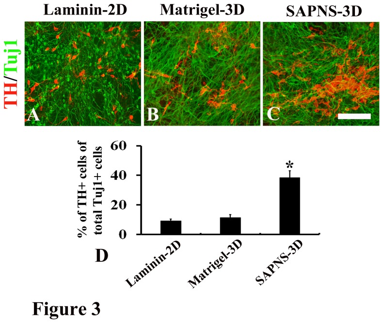
Dopaminergic differentiation of embryonic stem cells (ESCs) gains more and more attention worldwide owing to its potential use for neurorestorative therapy for the treatment of Parkinson's disease. The conventional 2D cell culture on petri dishes with various animal derived substrata such as collagen gels, laminin, and Matrigel is widely used to induce dopaminergic differentiation and it may limit the efficiency in the generation of dopaminergic neurons from ESCs and prevent their application for human therapies. Here, we reported that a self-assembling peptide made from natural amino acids has a property to generate a true 3D environment for dopaminergic differentiation. Mouse ESCs (R1) and mouse iPSCs (TTF-1) embedded in RADA16-I peptide-derived nanofiber scaffolds led to a marked increase in dopaminergic differentiation compared to the laminin-coated 2D culture or Matrigel-encapsulated 3D culture. These differentiated neurons expressed specific dopaminergic markers and produced appropriate patterns of action potential firing. Consistent with the increase in the number of dopaminergic neurons differentiated from R1 or TTF-1 in the self-assembling peptide nanofiber scaffold (SAPNS), both the expression levels of genes that involve in dopaminergic differentiation and maturation and the dopamine release in SAPNS culture were significantly elevated. The results of the study suggest that SAPNS provides a promising 3D culture system for dopaminergic differentiation.


Conflict of interest statement

Competing Interests: The authors have declared that no competing interests exist.

Figures

Figure 1
Figure 1. Derivation of neural progenitors from R1 and TTF-1 cells.
(A) and (D) Phase contrast image shows that R1 and TTF-1 cells grew as colonies on mitomycin-treated MEF feeder cells. (B) and (E) Phase contrast image shows neural rosettes derived from R1 and TTF-1 cells. (C) and (F) Immunostaining reveals that predominantly cells in rosettes R1 and TTF-1 cells were Nestin positive. Scale bar: 200 µm in A and D; 150 µm in B, C, E, and F.
Figure 2
Figure 2. Dopaminergic differentiation of R1 cells was significantly improved in SAPNS-3D culture.
(A) Immunocytochemistry for TH and Tuj1 revealed the dopaminergic differentiation of R1 in Laminin-2D culture. (B) Immunocytochemistry for TH and Tuj1 revealed the dopaminergic differentiation of R1 in Matrigel-3D culture. (C) Immunocytochemistry for TH and Tuj1 revealed the dopaminergic differentiation of R1 in SAPNS-3D culture. (D) The percentage of TH-positive cells of total Tuj1-positive cells was 41.5% ± 3.4% in SAPNS-3D culture, which was significantly higher than 8.3% ± 1.4% in the laminin-2D culture and 7.9% ± 1.6% in Matrigel-3D culture. *P<0.001. Scale bar: 150 µm.
Figure 3
Figure 3. Dopaminergic differentiation of TTF-1 cells was significantly improved in SAPNS-3D culture.
(A) Immunocytochemistry for TH and Tuj1 revealed the dopaminergic differentiation of TTF-1 in Laminin-2D culture. (B) Immunocytochemistry for TH and Tuj1 revealed the dopaminergic differentiation of TTF-1 in Matrigel-3D culture. (C) Immunocytochemistry for TH and Tuj1 revealed the dopaminergic differentiation of TTF-1 in SAPNS-3D culture. (D) The percentage of TH-positive cells of total Tuj1-positive cells was 38.5% ± 2.6% in SAPNS-3D culture, which was significantly higher than 9.2% ± 1.2% in the laminin-2D culture and 11.4% ± 2.1% in Matrigel-3D culture. *P<0.001. Scale bar: 150 µm.
Figure 4
Figure 4. The majority of TH positive neurons co-expressed specific midbrain dopaminergic neuron markers.
(A-C) Characterization of R1-derived TH positive neurons in SAPNS. (A) Double immunostaining showed that R1-derived TH positive neurons were co-labeled with Nurrl; (B) Double immunostaining showed that R1-derived TH positive neurons were co-labeled with Dat; (C) The majority of R1-derived TH positive neurons co-expressed Nurr1 or Dat. (D-F) Characterization of TTF-1-derived TH positive neurons in SAPNS. (D) Double immunostaining showed that TTF-1-derived TH positive neurons were co-labeled with Nurrl; (E) Double immunostaining showed that TTF-1-derived TH positive neurons were co-labeled with Dat; (F) The majority of TTF-1-derived TH positive neurons co-expressed Nurr1 or Dat. Scale bar: 100 µm.
Figure 5
Figure 5. Relative mRNA expression levels of dopaminergic differentiation markers (Lmx1a, Foxa2, En1, Aadc, Nurr1, Th, Vmat2, and Dat) in laminin-2D culture, Matrigel-3D culture and SAPNS-3D culture respectively.
Quantitative real-time RT-PCR demonstrated that a 4-fold increase for Lmx1a (A), 5-fold increase for Nurr1 (E), Vmat2 (G), and Dat (H), 6-fold increase for En1 (C) and Aadc (D), and 7-fold increase for Foxa2 (B) and Th (F) were found in both R1 and TTF-1 differentiation when cultured in SAPNS as compared to their expressions in Matrigel or on laminin.
Figure 6
Figure 6. Functional Analyses of dopaminergic neurons.
Standard whole-cell patch clamp, current-clamp techniques (A) and dopamine release measurement (B) were performed to explore the biological function of dopaminergic neurons. (A) Current injections (300 ms duration current injections with increasing 5 pA every round) and single current injection (300 ms duration, 20 pA) showing these dopaminergic neurons generated repetitive trains of action potentials. (B) HPLC analyses demonstrate that the dopamine level was increased more than 3-fold in SAPNS culture compared to that in Matrigel culture or laminin culture (* P<0.001 in both R1 and TTF-1 differentiation).

References

    1. Lee SH, Lumelsky N, Studer L, Auerbach JM, McKay RD (2000) Efficient generation of midbrain and hindbrain neurons from mouse embryonic stem cells. Nat Biotechnol 18: 675–679. doi:10.1038/76536. PubMed: 10835609. - DOI - PubMed
    1. Kawasaki H, Mizuseki K, Nishikawa S, Kaneko S, Kuwana Y et al. (2000) Induction of midbrain dopaminergic neurons from ES cells by stromal cell-derived inducing activity. Neuron 28: 31–40. doi:10.1016/S0896-6273(00)00083-0. PubMed: 11086981. - DOI - PubMed
    1. Perrier AL, Tabar V, Barberi T, Rubio ME, Bruses J et al. (2004) Derivation of midbrain dopamine neurons from human embryonic stem cells. Proc Natl Acad Sci U S A 101: 12543–12548. doi:10.1073/pnas.0404700101. PubMed: 15310843. - DOI - PMC - PubMed
    1. Schulz TC, Noggle SA, Palmarini GM, Weiler DA, Lyons IG et al. (2004) Differentiation of human embryonic stem cells to dopaminergic neurons in serum-free suspension culture. Stem Cells 22: 1218–1238. doi:10.1634/stemcells.2004-0114. PubMed: 15579641. - DOI - PubMed
    1. Zeng X, Cai J, Chen J, Luo Y, You ZB et al. (2004) Dopaminergic differentiation of human embryonic stem cells. Stem Cells 22: 925–940. doi:10.1634/stemcells.22-6-925. PubMed: 15536184. - DOI - PubMed

Publication types

LinkOut - more resources