

Adyuvant fractionated radiotherapy after resection of intracranial hemangiopericytoma
- PMID: 24377030
- PMCID: PMC3863145
- DOI: 10.1016/j.rpor.2012.03.005
Adyuvant fractionated radiotherapy after resection of intracranial hemangiopericytoma
Abstract
Aim: Review of literature and adjuvant treatment in Hemangiopericytoma after complete resection.
Background: Intracranial hemangiopericytoma (HPC) is an uncommon malignant vascular tumor arising from mesenchymal cells with pericytic differentiation. Surgery remains the mainstay treatment, and adjuvant radiation therapy appears to be appropriate for patients with high grade tumors or incomplete resection. We present our experience and review of the literature.
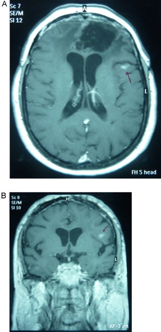
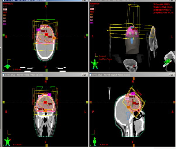
Materials and methods: We describe two cases of intracranial hemangiopericytoma located in the frontal lobe of the CNS. Both patients underwent complete tumor resection followed by adjuvant fractionated radiotherapy and completed treatment without interruptions.
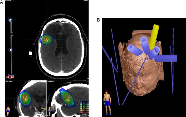
Results: A local recurrence was observed in one of these cases and fractionated stereotactic radiotherapy was performed. Both patients are alive and disease has been under control up to date.
Conclusion: The treatment of choice for intracranial hemangiopericytoma is a complete surgical resection as long as possible. Adjuvant radiotherapy of HPC can result in increased tumor control and should be considered as an effective treatment for patients with high grade or demonstrated residual tumor in the postoperative period. Salvage treatment using limited-field fractionated radiotherapy for local recurrence treatment is considered an acceptable option.
Keywords: Fractionated radiotherapy; Hemangiopericytoma; Radiotherapy; Stereotactic radiosurgery.
Figures


References
-
- Giannini C., Rushing E.J., Hainfellner J.A. Hemangiopericytoma. 3rd ed. 2007. Who classification of tumours of the central nervous system. pp. 178–180.
-
- Guthiere B.L., Ebersold M.J., Scheithauer B.W. Meningeal hemangiopericytoma: histopathological features, treatment, and long-term follow-up of 44 cases. Neurosurgery. 1989;25:514–522. - PubMed
-
- Jellinger K., Paulus W., Slowik F. The enigma of meningeal hemangiopericytoma. Brain Tumor Pathol. 1991;8:33–43.
-
- Fountas K.N., Kapsalaki E., Kassam M., Feltes C.H., Dimopoulos V.G., Robinson J.S., Smith J.R. Management of intracranial meningeal hemangiopericytomas: outcome and experience. Neurosurg Rev. 2006;29:145–153. - PubMed
-
- Sibtain N.A., Butt S., Connor S.E.J. Imaging features of central nervous system haemangiopericytomas. Eur Radiol. 2007;17:1685–1693. - PubMed
LinkOut - more resources
Full Text Sources
