

Recommendations of the Spanish Societies of Radiation Oncology (SEOR), Nuclear Medicine & Molecular Imaging (SEMNiM), and Medical Physics (SEFM) on (18)F-FDG PET-CT for radiotherapy treatment planning
- PMID: 24377032
- PMCID: PMC3863311
- DOI: 10.1016/j.rpor.2012.10.001
Recommendations of the Spanish Societies of Radiation Oncology (SEOR), Nuclear Medicine & Molecular Imaging (SEMNiM), and Medical Physics (SEFM) on (18)F-FDG PET-CT for radiotherapy treatment planning
Abstract
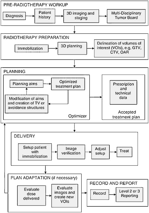
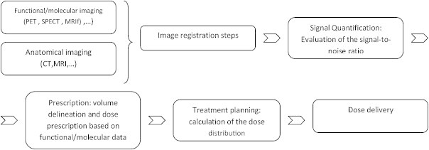
Positron emission tomography (PET) with (18)F-fluorodeoxyglucose (FDG) is a valuable tool for diagnosing and staging malignant lesions. The fusion of PET and computed tomography (CT) yields images that contain both metabolic and morphological information, which, taken together, have improved the diagnostic precision of PET in oncology. The main imaging modality for planning radiotherapy treatment is CT. However, PET-CT is an emerging modality for use in planning treatments because it allows for more accurate treatment volume definition. The use of PET-CT for treatment planning is highly complex, and protocols and standards for its use are still being developed. It seems probable that PET-CT will eventually replace current CT-based planning methods, but this will require a full understanding of the relevant technical aspects of PET-CT planning. The aim of the present document is to review these technical aspects and to provide recommendations for clinical use of this imaging modality in the radiotherapy planning process.
Keywords: CT; Contouring; Delineation; PET; PET-CT; Radiotherapy planning.
Figures

References
-
- Report 83. J ICRU. 2010;10(1) Oxford University Press. - PubMed
-
- Prescribing, recording and reporting photon beam therapy (supplement to ICRU report 50). ICRU report 62.
-
- Facey K., Bradbury I., Laking G., Payne E. Overview of the clinical effectiveness of positron emission tomography imaging in selected cancers. Health Technol Assess. 2007;11(October (44)) iii–iv, xi–267. - PubMed
-
- Delbeke D., Schöder H., Martin W.H., Wahl R.L. Hybrid imaging (SPECT/CT and PET/CT): improving therapeutic decisions. Semin Nucl Med. 2009;39(September (5)):308–340. - PubMed
LinkOut - more resources
Full Text Sources
Other Literature Sources
