. 2014 Jan 28;8(1):104-12.
doi: 10.1021/nn405701q. Epub 2014 Jan 7.

Gold-loaded polymeric micelles for computed tomography-guided radiation therapy treatment and radiosensitization

Affiliations

Gold-loaded polymeric micelles for computed tomography-guided radiation therapy treatment and radiosensitization

Ajlan Al Zaki et al. ACS Nano. .

Abstract

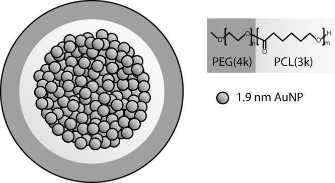
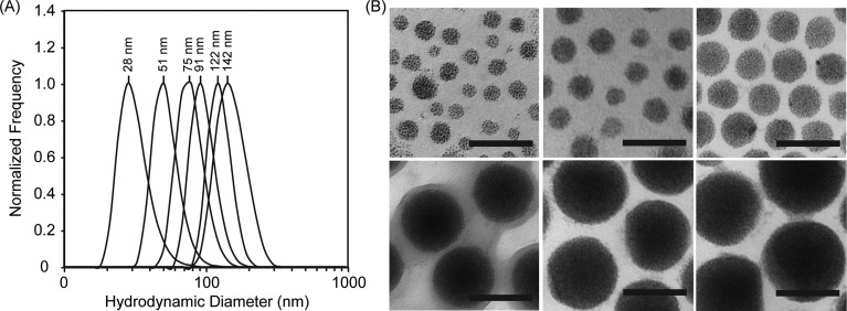
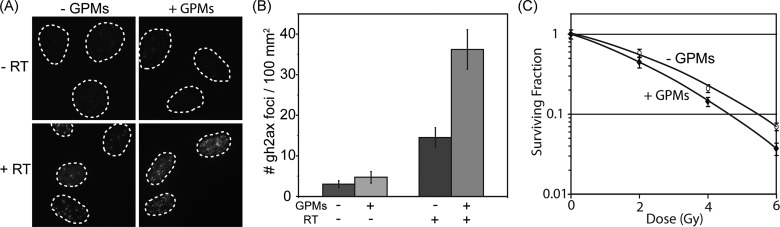
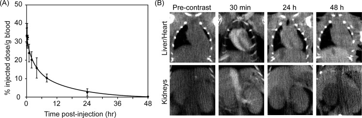
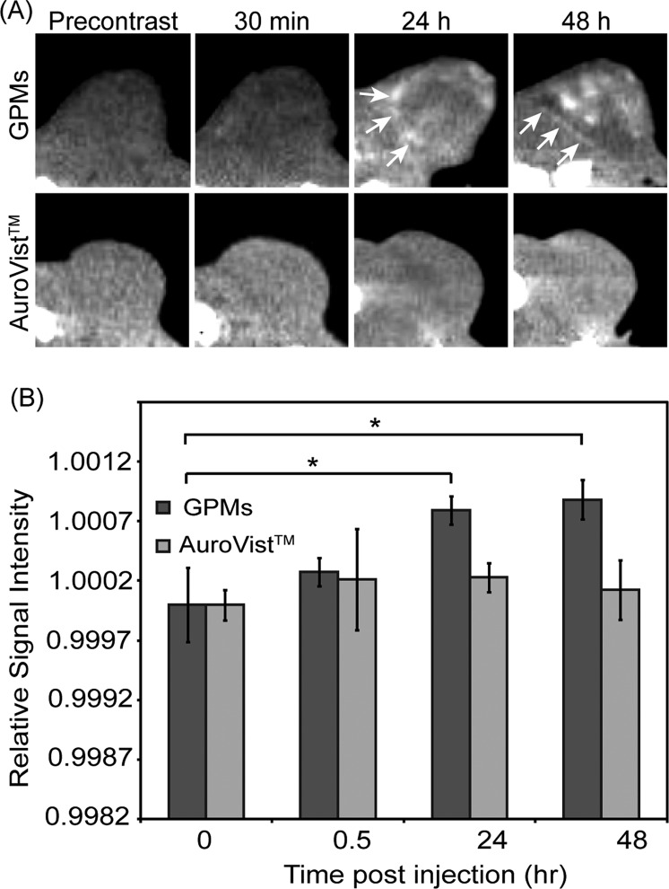
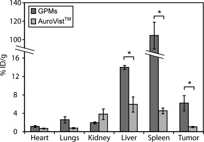
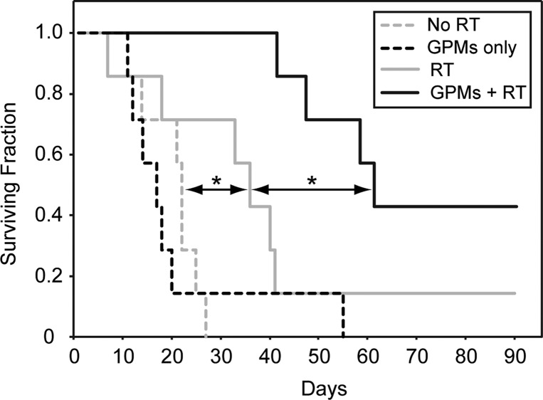
Gold nanoparticles (AuNPs) have generated interest as both imaging and therapeutic agents. AuNPs are attractive for imaging applications since they are nontoxic and provide nearly three times greater X-ray attenuation per unit weight than iodine. As therapeutic agents, AuNPs can sensitize tumor cells to ionizing radiation. To create a nanoplatform that could simultaneously exhibit long circulation times, achieve appreciable tumor accumulation, generate computed tomography (CT) image contrast, and serve as a radiosensitizer, gold-loaded polymeric micelles (GPMs) were prepared. Specifically, 1.9 nm AuNPs were encapsulated within the hydrophobic core of micelles formed with the amphiphilic diblock copolymer poly(ethylene glycol)-b-poly(ε-capralactone). GPMs were produced with low polydispersity and mean hydrodynamic diameters ranging from 25 to 150 nm. Following intravenous injection, GPMs provided blood pool contrast for up to 24 h and improved the delineation of tumor margins via CT. Thus, GPM-enhanced CT imaging was used to guide radiation therapy delivered via a small animal radiation research platform. In combination with the radiosensitizing capabilities of gold, tumor-bearing mice exhibited a 1.7-fold improvement in the median survival time, compared with mice receiving radiation alone. It is envisioned that translation of these capabilities to human cancer patients could guide and enhance the efficacy of radiation therapy.


Figures

Figure 1
Figure 1
Schematic of gold-loaded polymeric micelles (GPMs). Gold nanoparticles are self-assembled into the hydrophobic core of micelles, stabilized with the amphiphilic diblock copolymer PEG-b-PCL. Each GPM is composed of approximately hundreds to thousands of individual gold nanoparticles, depending on their size.
Figure 2
Figure 2
Size and morphology of GPMs. (A) Dynamic light scattering profiles of six GPM formulations with mean sizes ranging from 25 to 150 nm, in phosphate-buffered saline, pH 7.4. (B) Transmission electron microscopy (TEM) images of the same six GPM formulations, respectively. The electron micrographs reveal a narrow monodispersed distribution of spherical GPMs, with tightly packed gold clusters contained within the hydrophobic core (all scale bars = 100 nm).
Figure 3
Figure 3
In vitro evaluation of radiation-induced DNA double-strand breaks in the presence and absence of GPMs. (A) Immunofluorescent imaging of γ-h2ax foci in HT1080 cells incubated with or without GPMs in the absence (top) or presence (bottom) of irradiation (4 Gy). (B) Quantitative analysis of γ-h2ax foci density (# foci/μm2) for n > 100 cells in each treatment group. Error bars represent 95% confidence intervals. (C) Clonogenic assay of HT1080 cells treated with and without GPMs and given radiation doses of 0, 2, 4, and 6 Gy. Error bars represent the mean survival ± standard error of at least three replicates.
Figure 4
Figure 4
Blood clearance profile and CT imaging of GPMs in the blood pool. (A) ICP-OES analysis of gold content in blood at various times following the intravenous administration of GPMs to mice (n = 3). (B) Serial CT coronal views of a mouse following retro-orbital injection of 200 μL of GPM solution (650 mg/kg). Coronal views of heart and liver (top) and inferior vena cava and kidneys (bottom) are shown.
Figure 5
Figure 5
In vivo CT images and intensity analysis of nu/nu nude mice with HT1080 flank tumors. (a) Representative CT images in the axial plane prior to injection (precontrast) and 30 min, 24 h, and 48 h postinjection of GPMs (n = 3) or AuroVist (n = 3). Tumor boundaries are indicated by white arrows. (b) Quantitative analysis of CT images. Signal intensity of each tumor was normalized to adjacent paraspinal muscle. For contrast measurement, the relative signal intensity was calculated as the quotient of the postcontrast to precontrast normalized tumor intensity. The asterisk indicates statistical significance (p < 0.05).
Figure 6
Figure 6
ICP-OES analysis of gold distribution at 48 h following the administration of GPMs or AuroVist. The percent injected dose per gram of tissue was calculated by measuring the concentration of gold in excised organs via ICP-OES. The asterisk indicates statistical significance (p < 0.05).
Figure 7
Figure 7
Kaplan–Meier survival analysis. A survival analysis was performed for tumor-bearing mice (n = 7 per group) receiving no treatment (dotted gray line), GPMs only (dotted black line), irradiation only (solid gray line), or irradiation 24 h after retro-orbital injection of GPMs (solid black line). GPMs were administered at a dose of 650 mg Au/kg. The radiation dose administered was 6 Gy at 150 kVp. The asterisk indicates statistical significance (p < 0.05).

References

    1. Benita S.; Poly P. A.; Puisieux F.; Delattre J. Radiopaque Liposomes: Effect of Formulation Conditions on Encapsulation Efficiency. J. Pharm. Sci. 1984, 73, 1751–1755. - PubMed
    1. Elrod D. B.; Partha R.; Danila D.; Casscells S. W.; Conyers J. L. An Iodinated Liposomal Computed Tomographic Contrast Agent Prepared from a Diiodophosphatidylcholine Lipid. Nanomedicine 2009, 5, 42–45. - PubMed
    1. Sachse A.Iodinated Liposomes as Contrast Agents. In Fundamental Biomedical Technologies: Nanoparticles in Biomedical Imaging; Ferrari M., Bulte J. W. M., Modo M. M. J., Eds.; Springer: New York, 2008; pp 371–410.
    1. Torchilin V. P.; Frank-Kamenetsky M. D.; Wolf G. L. CT Visualization of Blood Pool in Rats by Using Long-Circulating, Iodine-Containing Micelles. Acad. Radiol. 1999, 6, 61–65. - PubMed
    1. Long D. M. Jr.; Lasser E. C.; Sharts C. M.; Multer F. K.; Nielsen M. Experiments with Radiopaque Perfluorocarbon Emulsions for Selective Opacification of Organs and Total Body Angiography. Invest. Radiol. 1980, 15, 242–247. - PubMed

Publication types