. 2013 Dec 30:11:122.
doi: 10.1186/1741-7007-11-122.

IpsA, a novel LacI-type regulator, is required for inositol-derived lipid formation in Corynebacteria and Mycobacteria

Affiliations

IpsA, a novel LacI-type regulator, is required for inositol-derived lipid formation in Corynebacteria and Mycobacteria

Meike Baumgart et al. BMC Biol. .

Abstract

Background: The development of new drugs against tuberculosis and diphtheria is focused on disrupting the biogenesis of the cell wall, the unique architecture of which confers resistance against current therapies. The enzymatic pathways involved in the synthesis of the cell wall by these pathogens are well understood, but the underlying regulatory mechanisms are largely unknown.

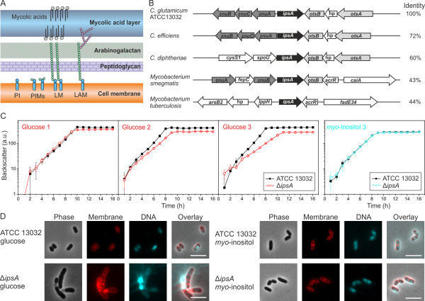
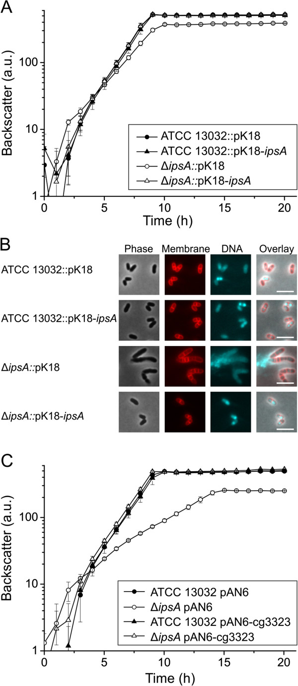
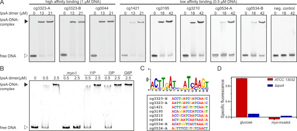
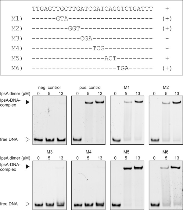
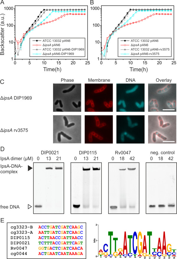
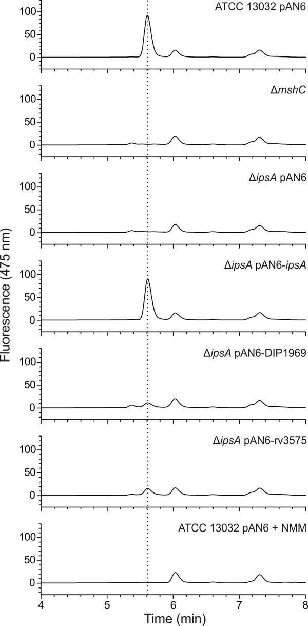
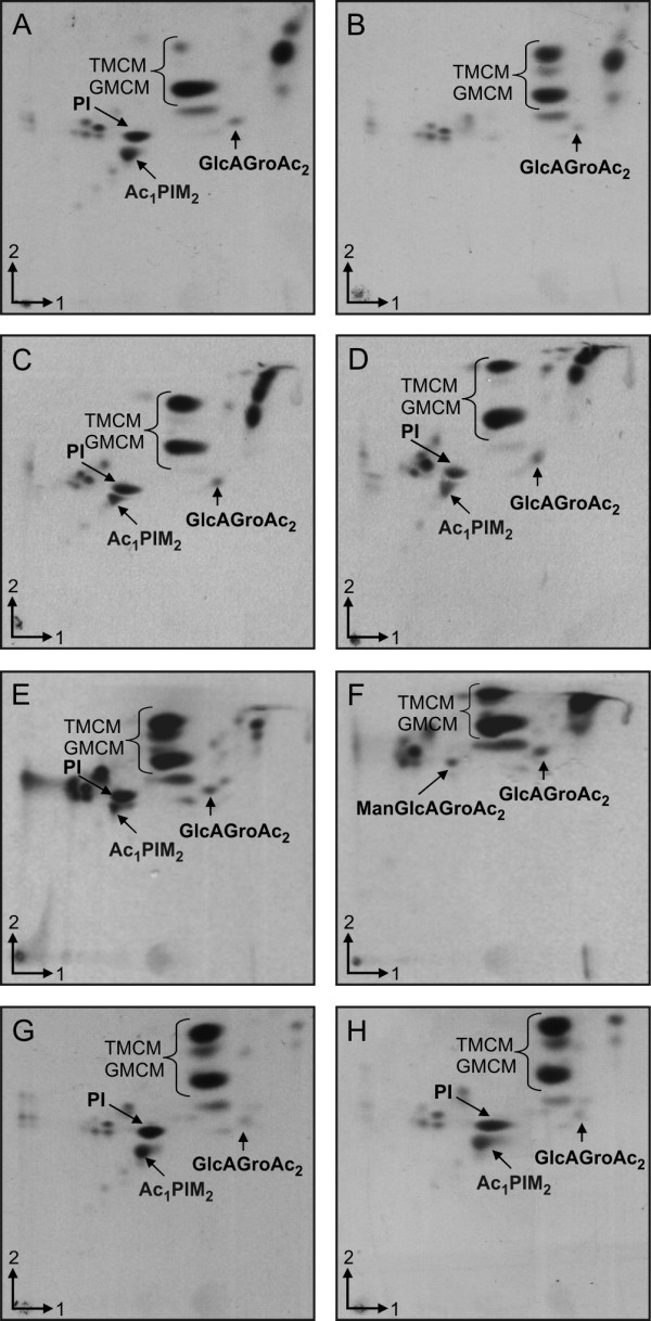
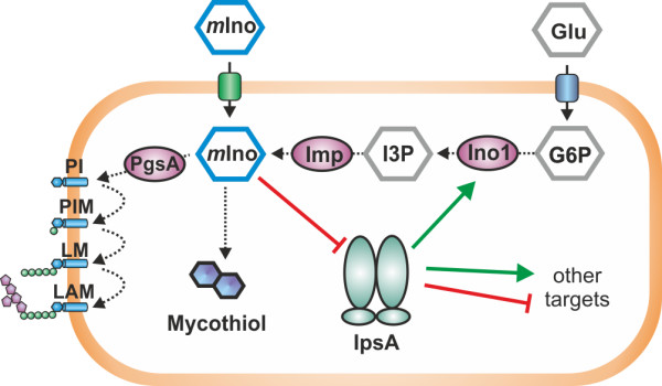
Results: Here, we characterize IpsA, a LacI-type transcriptional regulator conserved among Mycobacteria and Corynebacteria that plays a role in the regulation of cell wall biogenesis. IpsA triggers myo-inositol formation by activating ino1, which encodes inositol phosphate synthase. An ipsA deletion mutant of Corynebacterium glutamicum cultured on glucose displayed significantly impaired growth and presented an elongated cell morphology. Further studies revealed the absence of inositol-derived lipids in the cell wall and a complete loss of mycothiol biosynthesis. The phenotype of the C. glutamicum ipsA deletion mutant was complemented to different extend by homologs from Corynebacterium diphtheriae (dip1969) and Mycobacterium tuberculosis (rv3575), indicating the conserved function of IpsA in the pathogenic species. Additional targets of IpsA with putative functions in cell wall biogenesis were identified and IpsA was shown to bind to a conserved palindromic motif within the corresponding promoter regions. Myo-inositol was identified as an effector of IpsA, causing the dissociation of the IpsA-DNA complex in vitro.

Conclusions: This characterization of IpsA function and of its regulon sheds light on the complex transcriptional control of cell wall biogenesis in the mycolata taxon and generates novel targets for drug development.


Figures

Figure 1
Figure 1
IpsA plays an essential role in the integrity of the cell wall of Corynebacteriales. (A) Current model of the structure of the cell wall of Corynebacteria and Mycobacteria. (B) Comparison of the organization of the IpsA genome locus in C. glutamicum and related species. IpsA and homologous proteins are shown in black. Data were taken from [5,6]. The percentage identity of the amino acid sequences to IpsA from C. glutamicum was taken from NCBI Blast. hp, hypothetical protein. (C, D) Growth curves (C) and phenotype (D) of C. glutamicum wild-type and ΔipsA. Results in (C) show the growth (backscatter) from sequential cultures in minimal medium with glucose or myo-inositol as carbon source. After reaching the stationary phase, the cells were diluted into fresh medium at an initial OD600 of 1. Shown are three glucose cultures and the third myo-inositol culture (n = 3). In (D), DNA was stained with 4’,6-diamidino-2-phenylindole (DAPI) (cyan) and lipophilic regions with nile red (red). The scale bar is 5 μm.
Figure 2
Figure 2
Complementation of the ΔipsA phenotype with ipsA and the target gene ino1 (cg3323). For chromosomal complementation, the gene ipsA was integrated into the intergenic region of cg1121 to cg1122 under control of the native promoter. (A) Growth of the strains in CGXII minimal medium with 2% (w/v) glucose as carbon source. The average and standard deviation of three biological replicates is shown. (B) Fluorescence microscopy images of stationary phase cells of the cultures presented in (A). (C) Plasmid-based complementation with the target gene ino1 (cg3323). The strains were cultivated in CGXII minimal medium with 2% (w/v) glucose and 50 μM isopropyl β-d-1-thiogalactopyranoside (IPTG) for induction of ino1 expression.
Figure 3
Figure 3
Target genes, effector and binding sites of IpsA. (A) Binding of IpsA to 30 bp oligonucleotides covering the binding sites in the putative target promoters. As negative control the oligonucleotide cg3323-d (sequence AGGTCTGATTTCTGCTGGGAATCCCCACAT) was used, which is located immediately downstream of the IpsA binding sites in the cg3323 promoter. (B) The influence of intermediates in inositol metabolism (5 mM each) on the binding of IpsA to the ino1 promoter. myo-I, myo-inositol; I1P, 1D-myo-inositol-1-phosphate; I3P, 1D-myo-inositol-3-phosphate; G6P, glucose-6-phosphate. (C) The IpsA consensus motif predicted by MEME [20] and an overview of the sites in the corresponding promoter regions. (D) Promoter-fusion studies using the promoter of ino1 fused to eyfp (pJC1-Pcg3323-eyfp). The specific fluorescence of ATCC 13032 containing this plasmid on glucose was set to 1 and the other values were calculated accordingly.
Figure 4
Figure 4
Mutational analysis of one of the IpsA binding sites in the ino1 promoter. The importance of the predicted DNA sequence motif for IpsA binding was tested in electrophoretic mobility shift assays (EMSAs) with DNA fragments in which three nucleotides of the proposed motif were exchanged, as indicated. A + indicates that the mutated fragment was bound with the same affinity as the unaltered wild-type fragment (positive control); (+) indicates that the mutated fragment was shifted, but with lower affinity; - indicates that the mutated fragment was not shifted. In the case of M3 and M4, binding was completely abolished by the mutation, indicating that the six central base pairs are crucial for IpsA binding. For M1, M2 and M6 a slight decrease of binding was observed. The binding of M5 was unchanged.
Figure 5
Figure 5
IpsA in Corynebacterium diphtheriae and Mycobacterium tuberculosis.Corynebacterium glutamicum ΔipsA carrying the plasmids pAN6, pAN6-DIP1969 or pAN6-rv3573 and C. glutamicum wild-type with pAN6 as control were cultivated in CGXII with glucose without isopropyl β-d-1-thiogalactopyranoside (IPTG) (A) or with 50 μM IPTG (B). (C) Microscopic phenotypes of the complemented strains. DNA was stained with 4’,6-diamidino-2-phenylindole (DAPI) (cyan) and lipophilic regions with nile red (red), scale bar 5 μm. (D) Oligonucleotides (30 bp, 1 μM for DIP0021 and DIP0115, 0.5 μM for Rv0047 and the negative control) covering the predicted binding sites in the promoter regions of the respective genes were incubated with IpsA at the given concentrations and analyzed on 15% native polyacrylamide gels. In M. tuberculosis, Rv0046 is organized in an operon with Rv0047. A binding site was identified within the open reading frame (ORF) of Rv0047, suggesting the occurrence of a second promoter upstream of ino1. (E) IpsA binding motif derived from the high affinity C. glutamicum targets and the C. diphtheriae and M. tuberculosis binding sites.
Figure 6
Figure 6
Determination of mycothiol production in Corynebacterium glutamicum wild-type and different mutants and complemented strains. The mycothiol was derivatized using bromobimane, separated by high-performance liquid chromatography (HPLC) and monitored using a fluorescence detector (390 nm excitation and 475 nm emission). As control, the mycothiol deficient strain ΔmshC was used as well as a wild-type sample that had been treated with N-methyl-maleimide (NMM) to block thiols prior to derivatization. The peak assumed to be mycothiol is marked with a dotted line. Presented is a representative chromatogram of three biological replicates each.
Figure 7
Figure 7
Two-dimensional thin-layer chromatography (2D-TLC) analysis of [14C]-labeled polar lipids from Corynebacterium glutamicum cells grown in glucose (A-F) or myo-inositol (G-H). (A) and (G)C. glutamicum ATCC 13032, (B) and (H) ATCC 13032 ΔipsA,(C) ATCC 13032 ΔipsA pAN6-cg3323,(D) ATCC 13032 ΔipsA::pK18int-ipsA,(E) ATCC 13032 ΔipsA pAN6-DIP1969 and (F) ATCC 13032 ΔipsA pAN6-Rv3575. The cells were cultured in CGXII with either glucose (A-F) or myo-inositol (G, H). PI, phosphatidylinositol; TMCM, trehalose monocorynomycolate; GMCM, glucose monocorynomycolate; Ac1PIM2, monoacylated phosphatidyl myo-inositol dimannoside; GlcAGroAc2, 1,2-di-O-C16/C18:1-(α-d-glucopyranosyluronic acid)-(1→3)-glycerol (GL-A); ManGlcAGroAc2 1,2-di-O-C16/C18:1-(α-d-mannopyranosyl)-(1→4)-(α-d-glucopyranosyluronic acid)-(1→3)-glycerol (GL-X).
Figure 8
Figure 8
Model of IpsA function. In Corynebacteria and Mycobacteria, myo-inositol (mIno) is an important building block for cell wall components and mycothiol. mIno can be taken up from the culture medium or synthesized from glucose-6-phosphate (G6P) via 1D-myo-inositol-3-phosphate (I3P). When mIno concentration is low, IpsA activates ino1, encoding myo-inositol phosphate synthase, which catalyzes the formation of I3P from G6P. Besides ino1, IpsA activates or represses several other targets of unknown function.

References

    1. Mishra AK, Driessen NN, Appelmelk BJ, Besra GS. Lipoarabinomannan and related glycoconjugates: structure, biogenesis and role in Mycobacterium tuberculosis physiology and host-pathogen interaction. FEMS Microbiol Rev. 2011;11:1126–1157. doi: 10.1111/j.1574-6976.2011.00276.x. - DOI - PMC - PubMed
    1. Eggeling L, Besra GS, Alderwick L. In: Corynebacteria: genomics and molecular biology. Burkovski A, editor. Wymondham, UK: Caister Academic Press; 2008. Structure and synthesis of the cell wall; pp. 267–294.
    1. Fahey RC. Glutathione analogs in prokaryotes. Biochim Biophys Acta. 1830;11:3182–3198. - PubMed
    1. Morita YS, Fukuda T, Sena CBC, Yamaryo-Botte Y, McConville MJ, Kinoshita T. Inositol lipid metabolism in mycobacteria: biosynthesis and regulatory mechanisms. Biochim Biophys Acta. 1810;11:630–641. - PubMed
    1. http://www.microbesonline.org.

Publication types