

Testosterone dose dependently prevents bone and muscle loss in rodents after spinal cord injury
- PMID: 24378197
- PMCID: PMC5911705
- DOI: 10.1089/neu.2013.3155
Testosterone dose dependently prevents bone and muscle loss in rodents after spinal cord injury
Abstract
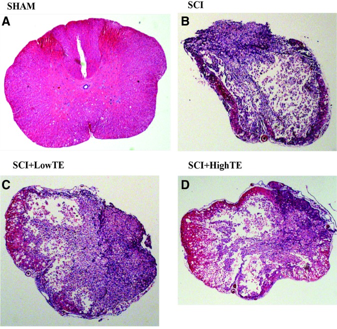
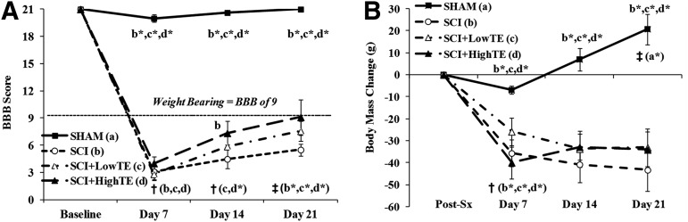
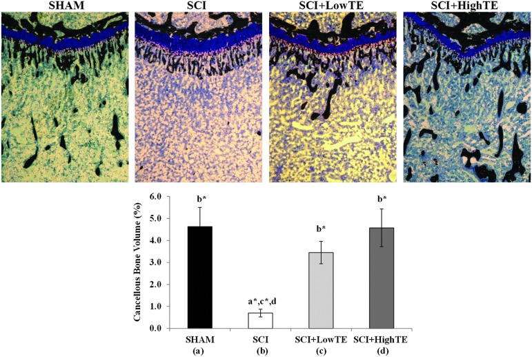
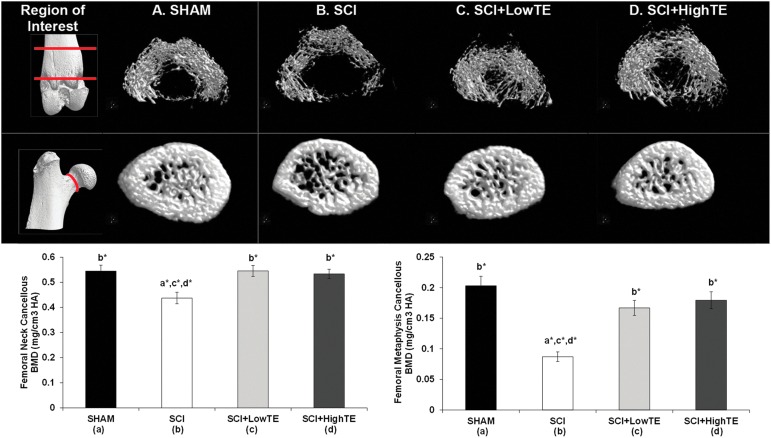
Androgen administration protects against musculoskeletal deficits in models of sex-steroid deficiency and injury/disuse. It remains unknown, however, whether testosterone prevents bone loss accompanying spinal cord injury (SCI), a condition that results in a near universal occurrence of osteoporosis. Our primary purpose was to determine whether testosterone-enanthate (TE) attenuates hindlimb bone loss in a rodent moderate/severe contusion SCI model. Forty (n=10/group), 14 week old male Sprague-Dawley rats were randomized to receive: (1) Sham surgery (T9 laminectomy), (2) moderate/severe (250 kdyne) SCI, (3) SCI+Low-dose TE (2.0 mg/week), or (4) SCI+High-dose TE (7.0 mg/week). Twenty-one days post-injury, SCI animals exhibited a 77-85% reduction in hindlimb cancellous bone volume at the distal femur (measured via μCT) and proximal tibia (measured via histomorphometry), characterized by a >70% reduction in trabecular number, 13-27% reduction in trabecular thickness, and increased trabecular separation. A 57% reduction in cancellous volumetric bone mineral density (vBMD) at the distal femur and a 20% reduction in vBMD at the femoral neck were also observed. TE dose dependently prevented hindlimb bone loss after SCI, with high-dose TE fully preserving cancellous bone structural characteristics and vBMD at all skeletal sites examined. Animals receiving SCI also exhibited a 35% reduction in hindlimb weight bearing (triceps surae) muscle mass and a 22% reduction in sublesional non-weight bearing (levator ani/bulbocavernosus [LABC]) muscle mass, and reduced prostate mass. Both TE doses fully preserved LABC mass, while only high-dose TE ameliorated hindlimb muscle losses. TE also dose dependently increased prostate mass. Our findings provide the first evidence indicating that high-dose TE fully prevents hindlimb cancellous bone loss and concomitantly ameliorates muscle loss after SCI, while low-dose TE produces much less profound musculoskeletal benefit. Testosterone-induced prostate enlargement, however, represents a potential barrier to the clinical implementation of high-dose TE as a means of preserving musculoskeletal tissue after SCI.
Conflict of interest statement
No competing financial interests exist.
Figures
Similar articles
-
Sclerostin inhibition prevents spinal cord injury-induced cancellous bone loss.J Bone Miner Res. 2015 Apr;30(4):681-9. doi: 10.1002/jbmr.2396. J Bone Miner Res. 2015. PMID: 25359699 Free PMC article.
-
Testosterone Plus Finasteride Prevents Bone Loss without Prostate Growth in a Rodent Spinal Cord Injury Model.J Neurotrauma. 2017 Nov 1;34(21):2972-2981. doi: 10.1089/neu.2016.4814. Epub 2017 Jun 5. J Neurotrauma. 2017. PMID: 28338402 Free PMC article.
-
Locomotor training with adjuvant testosterone preserves cancellous bone and promotes muscle plasticity in male rats after severe spinal cord injury.J Neurosci Res. 2020 May;98(5):843-868. doi: 10.1002/jnr.24564. Epub 2019 Dec 4. J Neurosci Res. 2020. PMID: 31797423 Free PMC article.
-
Activity-Based Physical Rehabilitation with Adjuvant Testosterone to Promote Neuromuscular Recovery after Spinal Cord Injury.Int J Mol Sci. 2018 Jun 7;19(6):1701. doi: 10.3390/ijms19061701. Int J Mol Sci. 2018. PMID: 29880749 Free PMC article. Review.
-
Bone and muscle loss after spinal cord injury: organ interactions.Ann N Y Acad Sci. 2010 Nov;1211:66-84. doi: 10.1111/j.1749-6632.2010.05806.x. Ann N Y Acad Sci. 2010. PMID: 21062296 Review.
Cited by
-
Evaluating Sex Steroid Hormone Neuroprotection in Spinal Cord Injury in Animal Models: Is It Promising in the Clinic?Biomedicines. 2024 Jul 4;12(7):1478. doi: 10.3390/biomedicines12071478. Biomedicines. 2024. PMID: 39062051 Free PMC article. Review.
-
Longitudinal Examination of Bone Loss in Male Rats After Moderate-Severe Contusion Spinal Cord Injury.Calcif Tissue Int. 2019 Jan;104(1):79-91. doi: 10.1007/s00223-018-0471-8. Epub 2018 Sep 14. Calcif Tissue Int. 2019. PMID: 30218117 Free PMC article.
-
Sclerostin inhibition prevents spinal cord injury-induced cancellous bone loss.J Bone Miner Res. 2015 Apr;30(4):681-9. doi: 10.1002/jbmr.2396. J Bone Miner Res. 2015. PMID: 25359699 Free PMC article.
-
Reactions of the rat musculoskeletal system to compressive spinal cord injury (SCI) and whole body vibration (WBV) therapy.J Musculoskelet Neuronal Interact. 2015 Jun;15(2):123-36. J Musculoskelet Neuronal Interact. 2015. PMID: 26032204 Free PMC article.
-
Wheelchair Tai Chi Ball Exercise for Improving Neuromuscular Functions of Older Adults With Disability.Front Aging Neurosci. 2022 Jul 19;14:935986. doi: 10.3389/fnagi.2022.935986. eCollection 2022. Front Aging Neurosci. 2022. PMID: 35928991 Free PMC article.
References
-
- Devivo M.J. (2012). Epidemiology of traumatic spinal cord injury: trends and future implications. Spinal Cord 50, 365–372 - PubMed
-
- Zehnder Y., Luthi M., Michel D., Knecht H., Perrelet R., Neto I., Kraenzlin M., Zach G., and Lippuner K. (2004). Long-term changes in bone metabolism, bone mineral density, quantitative ultrasound parameters, and fracture incidence after spinal cord injury: a cross-sectional observational study in 100 paraplegic men. Osteoporos. Int. 15, 180–189 - PubMed
-
- Eser P., Frotzler A., Zehnder Y., Wick L., Knecht H., Denoth J., and Schiessl H. (2004). Relationship between the duration of paralysis and bone structure: a pQCT study of spinal cord injured individuals. Bone 34, 869–880 - PubMed
-
- Frisbie J.H. (1997). Fractures after myelopathy: the risk quantified. J. Spinal Cord Med. 20, 66–69 - PubMed
Publication types
MeSH terms
Substances
Grants and funding
LinkOut - more resources
Full Text Sources
Other Literature Sources
Medical
