Review
. 2013 Dec 27;6(1):13-53.
doi: 10.3390/v6010013.

Replication cycle and molecular biology of the West Nile virus

Affiliations
Review

Replication cycle and molecular biology of the West Nile virus

Margo A Brinton. Viruses. .

Abstract

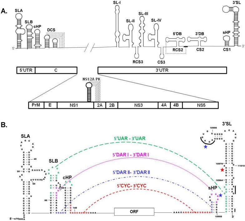
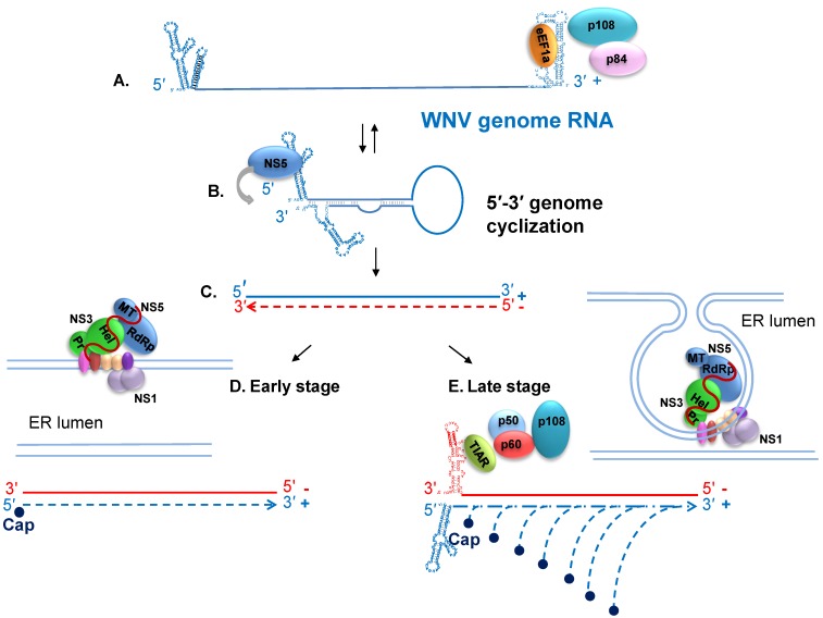
West Nile virus (WNV) is a member of the genus Flavivirus in the family Flaviviridae. Flaviviruses replicate in the cytoplasm of infected cells and modify the host cell environment. Although much has been learned about virion structure and virion-endosomal membrane fusion, the cell receptor(s) used have not been definitively identified and little is known about the early stages of the virus replication cycle. Members of the genus Flavivirus differ from members of the two other genera of the family by the lack of a genomic internal ribosomal entry sequence and the creation of invaginations in the ER membrane rather than double-membrane vesicles that are used as the sites of exponential genome synthesis. The WNV genome 3' and 5' sequences that form the long distance RNA-RNA interaction required for minus strand initiation have been identified and contact sites on the 5' RNA stem loop for NS5 have been mapped. Structures obtained for many of the viral proteins have provided information relevant to their functions. Viral nonstructural protein interactions are complex and some may occur only in infected cells. Although interactions between many cellular proteins and virus components have been identified, the functions of most of these interactions have not been delineated.


Figures

Figure 1
Figure 1
Schematic representations of the West Nile Virus (WNV) genomic RNA. (A) Conserved stem loop (SL) RNA structures located in the 3' and 5' untranslated regions (UTRs) as well as the capsid coding region are shown in the top panel. Pseudoknot interactions are indicated by dotted lines. The positions of the individual virus protein coding regions in the polyprotein ORF are shown in the bottom panel. Viral protein order from the N terminus of the polyprotein: capsid (C), pre-membrane (prM), envelop (E), and the nonstructural proteins, NS1 through NS5. A pseudoknot involved in a -1 translational frameshift that is required for the production of NS1' is also depicted in the lower panel; (B) Terminal region genome sequences that are involved in 3'–5' long distance RNA-RNA interactions. The interacting 5' upstream of the AUG (UAR) sequence and its 3' complement are indicated in green, the 5' downstream of the AUG 1 (DAR1) sequences in pink, the 5' downstream of the AUG 2 (DAR2) sequences in blue and the cyclization (CYC) sequences in orange. The black lines on the 3' SL indicate conserved sequences. The major binding site for eEF1A is indicated with a red star and the minor eEF1A binding sites are indicated by blue stars. Bracket, AUG of ORF.
Figure 2
Figure 2
Diagrams of WNV RNA replication at different phases of the viral replication cycle. (A) The linear form of the plus strand RNA genome is utilized as a template for translation; (B) The genome switches back and forth between linear and cyclized forms. The interaction of the cell protein eEF1A with three sites on the 3' stem loop (SL) RNA may facilitate the opening of 3' region basepairs prior to formation of the 3'-5' RNA-RNA interaction that cyclizes the genome. The cyclized form of the genome functions as the template for minus strand RNA synthesis. NS5 recruitment to the 3' end of the minus strand is facilitated by its interaction with the 5' SLA; (C) A single minus stand is copied from the genome; (D) Release of the 3' end of the minus strand from the template-product duplex by an unknown mechanism leads to initiation of the synthesis of a plus strand (genome) RNA. The viral replication complexes are associated with ER membranes at early stages of replication but ER invaginations are not yet observed and reinitiation of plus strand RNA synthesis is inefficient; (E) At later stages of the replication cycle, viral nonstructural proteins have induced the formation of invaginations in the ER membrane that contain viral replication complexes, cell proteins and minus strand templates. Late stage reinitiation of plus strand synthesis is very efficient. The complex formed by TIAR and other cell proteins and the 3' terminal SL of the minus strand is thought to enhance recruitment and possibly also the positioning of NS5 for plus strand reinitiation. Viral nonstructural protein: NS1 (medium blue), NS2A (dark red), NS2B (pink), NS3 (green), NS4A dimer (peach), and NS4B (purple).

References

    1. Berthet F.X., Zeller H.G., Drouet M.T., Rauzier J., Digoutte J.P., Deubel V. Extensive nucleotide changes and deletions within the envelope glycoprotein gene of Euro-African West Nile viruses. J. Gen. Virol. 1997;78:2293–2297. - PubMed
    1. Jia X.Y., Briese T., Jordan I., Rambaut A., Chi H.C., Mackenzie J.S., Hall R.A., Scherret J., Lipkin W.I. Genetic analysis of West Nile New York 1999 encephalitis virus. Lancet. 1999;354:1971–1972. doi: 10.1016/S0140-6736(99)05384-2. - DOI - PubMed
    1. Lanciotti R.S., Roehrig J.T., Deubel V., Smith J., Parker M., Steele K., Crise B., Volpe K.E., Crabtree M.B., Scherret J.H., et al. Origin of the West Nile virus responsible for an outbreak of encephalitis in the northeastern United States. Science. 1999;286:2333–2337. doi: 10.1126/science.286.5448.2333. - DOI - PubMed
    1. Beasley D.W., Li L., Suderman M.T., Barrett A.D. Mouse neuroinvasive phenotype of West Nile virus strains varies depending upon virus genotype. Virology. 2002;296:17–23. doi: 10.1006/viro.2002.1372. - DOI - PubMed
    1. Heinz F.X., Purcell M.S., Gould E.A., Howard C.R., Houghton M., Moormann R.J.M., Rice C.M., Thiel H.-J. Family Flaviviridae. In: Regenmortel C.F., Bishop D.H.L., Carstens E.B., Estes M.K., editors. Virus Taxonomy. Academic Press; San Diego, CA, USA: 2000. pp. 860–878.

Publication types

MeSH terms