Case Reports
doi: 10.11622/smedj.2013253.
Hirayama disease in a 17-year-old Chinese man
- PMID: 24379115
- PMCID: PMC4294056
- DOI: 10.11622/smedj.2013253
Item in Clipboard
Case Reports
Hirayama disease in a 17-year-old Chinese man
Singapore Med J.
2014 Jun.
Abstract
Hirayama disease is an uncommon cervical myelopathy associated with neck flexion. It has been postulated to be related to the anterior shifting of the posterior dura of the lower cervical dural canal during neck flexion, resulting in lower cervical cord atrophy with asymmetric flattening. We report a case of Hirayama disease in a 17-year-old Chinese man and demonstrate the use of dynamic flexion magnetic resonance imaging of the cervical spine in the diagnosis of the disease.
Figures
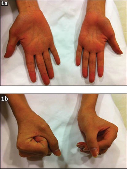
Photographs show (a) marked atrophy of the thenar, hypothenar and interosseous muscles of both hands, but more severely affecting the left side, and (b) impairment of the palmar grasp.
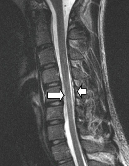
Non-flexion cervical T2-W sagittal MR image shows cord atrophy at the C5-C6 level (arrow), predominantly affecting the bilateral anterior aspects, and the posterior dura to be detached (arrowhead).
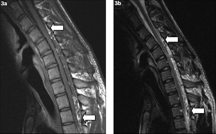
Flexion (a) T1-W and (b) T2-W sagittal MR images show anterior displacement of the posterior wall of the cervical dural canal from levels C4 to T3 (arrows), with flattening of the cord and complete effacement of the anterior thecal sac.
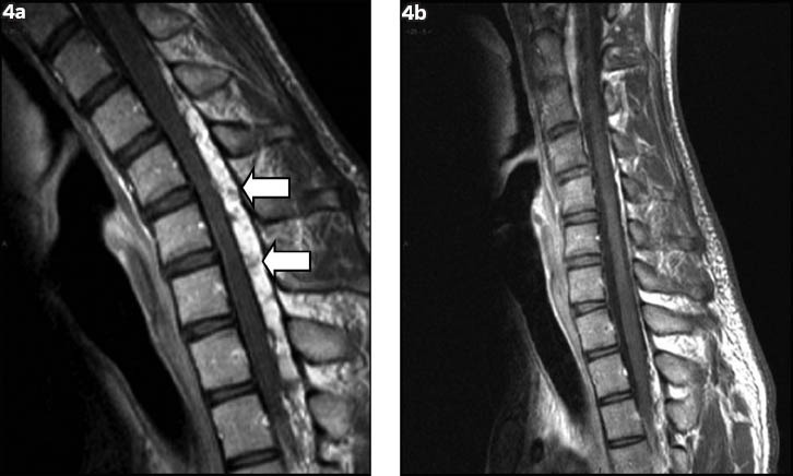
Dynamic T1-W postcontrast sagittal flexion MR images show (a) a homogeneously contrast-enhancing posterior epidural mass with multiple small signal voids within (arrows); and (b) disappearance of the epidural mass when the neck returns to a neutral position.
References
-
- Hirayama K, Toyokura Y, Tsubaki T. Juvenile muscular atrophy of unilateral upper extremity: a new clinical entity. Psychiatr Neurol Jpn. 1959;61:2190–7. - PubMed
-
- Pradhan S, Gupta RK. Magnetic resonance imaging in juvenile asymmetric segmental spinal muscular atrophy. J Neurol Sci. 1997;146:133–8. - PubMed
Publication types
MeSH terms
Supplementary concepts
LinkOut - more resources
Full Text Sources
Other Literature Sources