

The isolation and cultivation of bone marrow stem cells and evaluation of differences for neural-like cells differentiation under the induction with neurotrophic factors
- PMID: 24379142
- PMCID: PMC4235946
- DOI: 10.1007/s10616-013-9654-3
The isolation and cultivation of bone marrow stem cells and evaluation of differences for neural-like cells differentiation under the induction with neurotrophic factors
Abstract
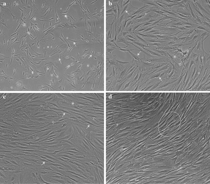
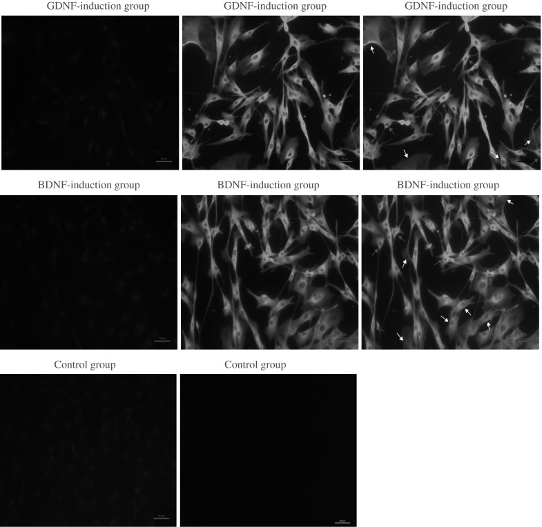
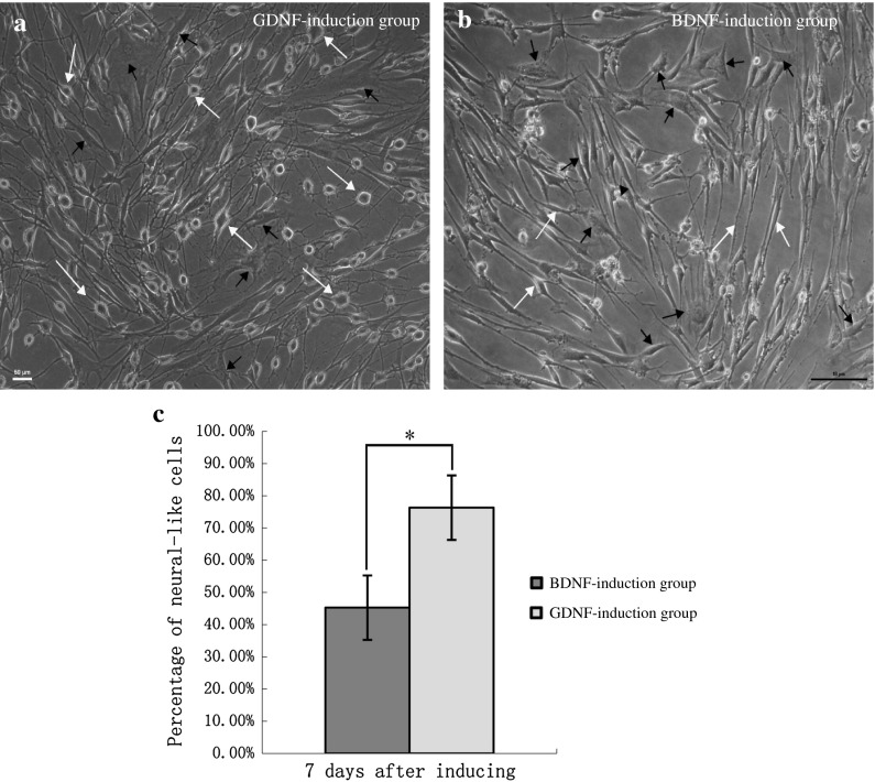
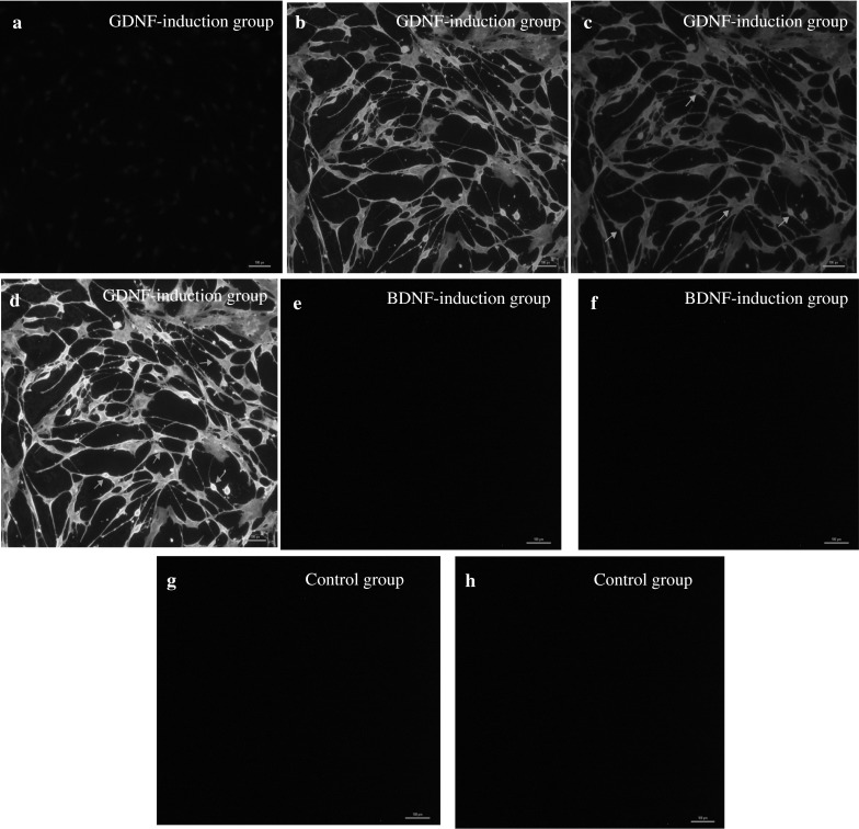
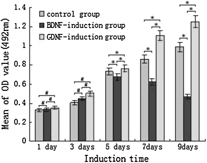
The bone marrow represents the most common source from which to isolate mesenchymal stem cells (MSCs). They can be obtained directly from patients and successfully induced to form various differentiated cell types. In addition, cell-based transplantation therapies have been proven to be promising strategies for curing disease of the nerve system. Therefore, it was particularly important to establish an easy and feasible method for the isolation, purification, and differentiation of bone marrow stromal cells (BMSCs). The aim of this study was to isolate and characterize putative bone marrow derived MSCs from Sprague-Dawley (SD) rats. Furthermore, differentiation effects were compared between the GDNF-induction group and the BDNF-induction group. Of these, BMSCs were isolated from the SD rats in a traditional manner, and identified based on plastic adherence, morphology, and surface phenotype assays. After induction with GDNF and BDNF, viability of BMSCs was detected by MTT assay and neuronal differentiation of BMSCs was confirmed by using immunofluorescence and Western blotting. Besides, the number of BMSCs that obviously exhibited neuronal morphology was counted and the results were compared between the GDNF-induction group and BDNF-induction groups. Our results indicate that direct adherence was a simple and convenient method for isolation and cultivation of BMSCs. Furthermore, BMSCs can be induced in vitro to differentiate into neuronal cells by using GDNF, which could achieve a more persistent and stable inducing effect than when using BDNF.
Figures


Similar articles
-
Bone marrow-derived mesenchymal stem cells differentiate into nerve-like cells in vitro after transfection with brain-derived neurotrophic factor gene.In Vitro Cell Dev Biol Anim. 2015 Mar;51(3):319-27. doi: 10.1007/s11626-015-9875-1. Epub 2015 Mar 14. In Vitro Cell Dev Biol Anim. 2015. PMID: 25773996 Free PMC article.
-
Improved stem cell therapy of spinal cord injury using GDNF-overexpressed bone marrow stem cells in a rat model.Biologicals. 2017 Nov;50:73-80. doi: 10.1016/j.biologicals.2017.08.009. Epub 2017 Aug 26. Biologicals. 2017. PMID: 28851622
-
In vitro neural differentiation of bone marrow stromal cells induced by hepatocyte growth factor and glial cell derived neurotrophic factor.Eur Rev Med Pharmacol Sci. 2016 Nov;20(22):4654-4663. Eur Rev Med Pharmacol Sci. 2016. PMID: 27906439
-
Glial cell induced neural differentiation of bone marrow stromal cells.Open Med (Wars). 2020 Sep 30;15(1):954-961. doi: 10.1515/med-2020-0229. eCollection 2020. Open Med (Wars). 2020. PMID: 33336053 Free PMC article.
-
Differentiation of GDNF and NT-3 dual gene-modified rat bone marrow mesenchymal stem cells into enteric neuron-like cells.J Huazhong Univ Sci Technolog Med Sci. 2012 Feb;32(1):87-91. doi: 10.1007/s11596-012-0015-9. Epub 2012 Jan 27. J Huazhong Univ Sci Technolog Med Sci. 2012. PMID: 22282251
Cited by
-
Visual bone marrow mesenchymal stem cell transplantation in the repair of spinal cord injury.Neural Regen Res. 2015 Mar;10(3):404-11. doi: 10.4103/1673-5374.153688. Neural Regen Res. 2015. PMID: 25878588 Free PMC article.
-
Donor mesenchymal stem cell-derived neural-like cells transdifferentiate into myelin-forming cells and promote axon regeneration in rat spinal cord transection.Stem Cell Res Ther. 2015 May 27;6(1):105. doi: 10.1186/s13287-015-0100-7. Stem Cell Res Ther. 2015. PMID: 26012641 Free PMC article.
-
Chitosan-collagen porous scaffold and bone marrow mesenchymal stem cell transplantation for ischemic stroke.Neural Regen Res. 2015 Sep;10(9):1421-6. doi: 10.4103/1673-5374.163466. Neural Regen Res. 2015. PMID: 26604902 Free PMC article.
-
Bone marrow-derived cells and their conditioned medium induce microvascular repair in uremic rats by stimulation of endogenous repair mechanisms.Sci Rep. 2017 Aug 25;7(1):9444. doi: 10.1038/s41598-017-09883-x. Sci Rep. 2017. PMID: 28842629 Free PMC article.
-
Recent advances of stem cell therapy for retinitis pigmentosa.Int J Mol Sci. 2014 Aug 20;15(8):14456-74. doi: 10.3390/ijms150814456. Int J Mol Sci. 2014. PMID: 25141102 Free PMC article. Review.
References
-
- Amemori T, Romanyuk N, Jendelova P, Herynek V, Turnovcova K, Prochazka P, Kapcalova M, Cocks G, Price J, Sykova E. Human conditionally immortalized neural stem cells improve locomotor function after spinal cord injury in the rat. Stem Cell Res Ther. 2013;4:68–83. doi: 10.1186/scrt219. - DOI - PMC - PubMed
-
- Bae JS, Han HS, Youn DH, Carter JE, Modo M, Schuchman EH, Jin HK (2007) Bone marrow-derived mesenchymal stem cells promote neuronal networks with functional synaptic transmission after transplantation into mice with neurodegeneration. Stem Cells 5:1307–1316 - PubMed
LinkOut - more resources
Full Text Sources
Other Literature Sources
