Review
. 2013 Dec 30;8(2):a016824.
doi: 10.1101/cshperspect.a016824.

Synaptic Signaling in Learning and Memory

Affiliations
Review

Synaptic Signaling in Learning and Memory

Mary B Kennedy. Cold Spring Harb Perspect Biol. .

Abstract

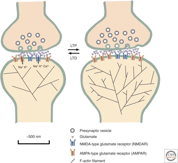
Learning and memory require the formation of new neural networks in the brain. A key mechanism underlying this process is synaptic plasticity at excitatory synapses, which connect neurons into networks. Excitatory synaptic transmission happens when glutamate, the excitatory neurotransmitter, activates receptors on the postsynaptic neuron. Synaptic plasticity is a higher-level process in which the strength of excitatory synapses is altered in response to the pattern of activity at the synapse. It is initiated in the postsynaptic compartment, where the precise pattern of influx of calcium through activated glutamate receptors leads either to the addition of new receptors and enlargement of the synapse (long-term potentiation) or the removal of receptors and shrinkage of the synapse (long-term depression). Calcium/calmodulin-regulated enzymes and small GTPases collaborate to control this highly tuned mechanism.


Figures

Figure 1.
Figure 1.
Features of excitatory neurons in the brain. The cell body (soma) of a typical excitatory pyramidal neuron is ∼10 μm in diameter and is located in one of several sheets of tightly packed somas that define the layers of the neocortex and hippocampus. Apical and basal dendrites extend from the soma, reaching into adjacent areas that are referred to as neuropil. Postsynaptic structures are located in tiny membrane protuberances called spines that can be seen along the dendrites. Each soma gives rise to one axon, which has a smaller diameter than the numerous dendrites. The axon can extend for millimeters from the soma and branches to form thousands of presynaptic terminals where transmitter is released onto the postsynaptic sites of other neurons. The axon hillock is located at the base of the axon. Action potentials are usually initiated at this site; they travel along the axon (arrows) to presynaptic terminals keeping a uniform amplitude of depolarization. Back-propagating action potentials travel in the opposite direction through the soma and into the dendrites. The size of their depolarization decreases as they travel and is regulated by the composition of dendritic ion channels. (From Peters and Kaiserman-Abramof 1970; modified, with permission, © Wiley.)
Figure 2.
Figure 2.
Synaptic plasticity. At the cellular level, one of the most essential elements of memory formation is the adjustment in synaptic strength of excitatory synapses between neurons. AMPA-type glutamate receptors (yellow) allow passage of sodium and potassium through their channel. Their principal function is to depolarize the membrane, producing an excitatory postsynaptic potential (EPSP). NMDA-type glutamate receptors (blue) also depolarize the membrane, but in addition to sodium and potassium, calcium flows through their channel and can initiate synaptic plasticity. A long-lasting increase in synaptic strength is referred to as long-term potentiation (LTP). LTP involves the addition of new synaptic AMPA-type glutamate receptors (AMPARs) and an increase in the size of the head of the postsynaptic spine, supported by an increase in the size and branching of the actin cytoskeleton. Long-term depression (LTD) is a long-lasting decrease in synaptic strength that involves a decrease in the number of synaptic AMPARs and shrinkage of the spine head. LTP is induced when repeated firing of an action potential in the presynaptic terminal and the resulting release of glutamate cause firing of action potentials in the postsynaptic neuron. LTD is induced when repeated firing of an action potential in the presynaptic terminal does not cause firing of action potentials in the postsynaptic neuron.
Figure 3.
Figure 3.
CaMKII. CaMKII is a ring of six dimers of calcium-/calmodulin (CaM)-activated catalytic subunits. The subunits are bound together by a central “hub” structure (light orange) formed from the carboxy-terminal association domains of each subunit. The inactive dimers (light and dark blue) are docked against the central hub by interactions among helices in the association domains (red) and residues in their inhibitory domains (light yellow). Binding of activated calmodulin to the subunit dimers is cooperative because binding to one subunit dissociates the dimer and makes the other subunit more available for calmodulin binding. The activated subunits are mobile and, in addition to phosphorylating other synaptic proteins, they can autophosphorylate each other at a critical threonine residue that locks the subunit in an active state until it is dephosphorylated by phosphatase 1 or phosphatase 2A. (From Rosenberg et al. 2005; modified, with permission, © Elsevier.)
Figure 4.
Figure 4.
Schematic diagram of the postsynaptic density (PSD) scaffold. NMDARs are immobilized in the postsynaptic membrane by association with PSD95. AMPARs associate with TARPs that bind PSD95. Signaling enzymes are positioned near the receptors by association with PSD95 or other scaffolds. For example, PKA, PKC, and calcineurin bind to AKAP79, which binds to PSD95 and actin in the spine. AMPARs are recruited to the PSD during induction of LTP by diffusional trapping at new docking sites. This diffusional trapping may involve phosphorylation of TARPs by CaMKII, which increases their binding affinity for PSD95. AMPARs are added to or removed from the extrasynaptic plasma membrane pool by exocytosis or endocytosis, respectively. (Modified from Sheng and Kim 2011.)
Figure 5.
Figure 5.
Signaling pathways in the spine. (A) Schematic diagram of major signaling enzymes that mediate changes in synaptic plasticity driven by calcium influx through activated NMDARs. Critical early targets of CaMKII include AMPARs, TARPs, and SynGAP. RasGRF can activate both Ras and Rac. The critical targets of calcineurin are not known. One possibility is the cofilin phosphatase Slingshot, which dephosphorylates and activates the F-actin regulator cofilin. Calcium/CaM-dependent AC and cAMP-phosphodiesterase (PDE) are both present in spines; their responses to calcium could generate a transient spike in cAMP, activating Epac. Ras and Rap regulate trafficking of AMPARs, but their downstream targets, beyond the MAPKs, are not yet known. RasGRF and Ras, acting through the GTPase exchange factor TIAM1 can activate Rac, which regulates pathways that control polymerization of actin. TrkB receptors that respond to the central nervous system (CNS) hormone brain-derived neural factor (BDNF) can provide tonic activation of Ras and Rap. (B) Synaptic regulation of AMPAR trafficking. A critical step in the induction of LTP is the trapping of additional AMPARs in the PSD scaffold through association of TARP and PSD95, which is increased by phosphorylation of residues on TARP by CaMKII. Dephosphorylation of these residues by PP1 can produce loss of AMPARs and depotentiation. Calcineurin, a calcium/CaM-dependent phosphatase, regulates PP1 and endocytosis. Addition of AMPARs to the dendritic plasma membrane by exocytosis, and their removal by endocytosis, occurs at perisynaptic sites in the spine and along the dendritic shaft. Changes in the activity of Ras and Rap are regulated by downstream targets of calcium/CaM, including RasGRF, SynGAP, and adenylyl cyclase (AC). Active Ras and Rap, in turn, activate the MAPKs ERK1/2 and p38. Pathways downstream from ERK facilitate exocytosis, whereas those downstream from p38 facilitate endocytosis. Many of the intermediate steps in the processes downstream from MAPKs are unknown.

References

    1. Abeliovich A, Chen C, Goda Y, Silva AJ, Stevens CF, Tonegawa S. 1993. Modified hippocampal long-term potentiation in PKCγ-mutant mice. Cell 75: 1253–1262. - PubMed
    1. Abraham WC, Bear MF. 1996. Metaplasticity: The plasticity of synaptic plasticity. Trends Neurosci 19: 126–130. - PubMed
    1. Allen PB, Hvalby O, Jensen V, Errington ML, Ramsay M, Chaudhry FA, Bliss TV, Storm-Mathisen J, Morris RG, Andersen P, et al. 2000. Protein phosphatase-1 regulation in the induction of long-term potentiation: Heterogeneous molecular mechanisms. J Neurosci 20: 3537–3543. - PMC - PubMed
    1. Ascher P, Nowak L. 1988. The role of divalent cations in the N-methyl-d-aspartate responses of mouse central neurones in culture. J Physiol 399: 247–266. - PMC - PubMed
    1. Bennett MK, Erondu NE, Kennedy MB. 1983. Purification and characterization of a calmodulin-dependent protein kinase that is highly concentrated in brain. J Biol Chem 258: 12735–12744. - PubMed

LinkOut - more resources