. 2014 Jan 28;111(4):1373-8.
doi: 10.1073/pnas.1318001111. Epub 2013 Dec 30.

Structural mechanisms of PriA-mediated DNA replication restart

Affiliations

Structural mechanisms of PriA-mediated DNA replication restart

Basudeb Bhattacharyya et al. Proc Natl Acad Sci U S A. .

Abstract

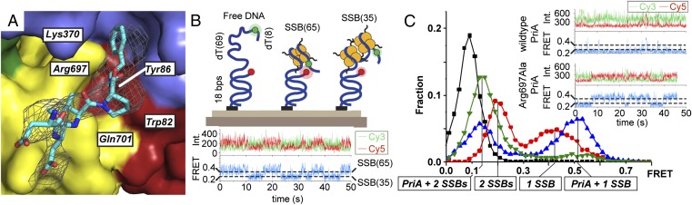
Collisions between cellular DNA replication machinery (replisomes) and damaged DNA or immovable protein complexes can dissociate replisomes before the completion of replication. This potentially lethal problem is resolved by cellular "replication restart" reactions that recognize the structures of prematurely abandoned replication forks and mediate replisomal reloading. In bacteria, this essential activity is orchestrated by the PriA DNA helicase, which identifies replication forks via structure-specific DNA binding and interactions with fork-associated ssDNA-binding proteins (SSBs). However, the mechanisms by which PriA binds replication fork DNA and coordinates subsequent replication restart reactions have remained unclear due to the dearth of high-resolution structural information available for the protein. Here, we describe the crystal structures of full-length PriA and PriA bound to SSB. The structures reveal a modular arrangement for PriA in which several DNA-binding domains surround its helicase core in a manner that appears to be poised for binding to branched replication fork DNA structures while simultaneously allowing complex formation with SSB. PriA interaction with SSB is shown to modulate SSB/DNA complexes in a manner that exposes a potential replication initiation site. From these observations, a model emerges to explain how PriA links recognition of diverse replication forks to replication restart.

Keywords: X-ray crystal structure; single-molecule FRET.


Conflict of interest statement

J.L.K. is a cofounder of Replisoma, Inc.

Figures

Fig. 1.
Fig. 1.
Structure of PriA DNA helicase. (A) Schematic diagram of PriA domain structure. (B) Crystal structure of KpPriA. Domains are colored as in A. ADP (red sticks) is bound within the helicase core, and two Zn2+ ions (gray spheres) are bound to the CRR. (C) Electrostatic surface features of KpPriA (blue, electropositive; red, electronegative; white, neutral). (D) Evolutionary conservation of KpPriA (a conservation scale, from variable to invariant among 150 PriA protein sequences, is shown below the structure).
Fig. 2.
Fig. 2.
PriA domain features. (A) Core helicase motifs and the ARL (magenta, labeled) line the interface between the bilobed HD. An ADP molecule (red sticks) is shown. (B) PriA CRR binds two Zn2+ ions (gray spheres) and positions a β-hairpin that is proposed to act as a DNA unwinding wedge. (C) Close-up view of the CTD (yellow) with basic residues displayed as sticks. The surfaces of the 3′BD, helicase core, and CRR are rendered to highlight the extensive contacts made between the CTD and each domain. (D) Overlay of KpPriA CTD (yellow) with the S10 ribosomal subunit (gray, with rRNA shown in orange) (37). RNA-binding basic residues from S10 and similarly positioned residues from the KpPriA CTD are shown (sticks). (E) EcPriA CTD binding to fluorescently labeled dT28 (black), 10-bp duplex (blue), 38-bp duplex (green), and replication fork (red) DNA. Data are the mean of three independent experiments, with error bars representing 1 SD.
Fig. 3.
Fig. 3.
PriA/SSB complex structure and function. (A) Fo-Fc omit electron density (contoured at 1.7-σ in the image) identifies the SSB-Ct peptide-binding site on PriA. Invariant PriA residues are labeled. (B, Upper) Schematic of the smFRET assay that distinguishes free DNA, SSB65-bound DNA, and SSB35-bound DNA (44). (B, Lower) Trace of an individual DNA molecule transitioning between SSB65- and SSB35-binding modes. Cy3 (green), Cy5 (red), and FRET (blue) intensities fluctuate over time due to mode interconversion. (C) smFRET-efficiency histograms of DNA alone (black), DNA with 20 nM SSB (red), 20 nM SSB with 1 μM PriA (green), and 20 nM SSB with 1 μM Arg697Ala PriA (blue). (Inset) Representative single-molecule time traces are shown for PriA and Arg697Ala PriA experiments. Int., intensity.
Fig. 4.
Fig. 4.
PriA replication restart initiation models. PriA recognizes abandoned DNA replication forks with either duplex (Upper) or SSB-bound ssDNA (Lower) lagging strands and processes these to expose ssDNA necessary for full primosome assembly and reloading of the replicative helicase. Nascent DNA strands are colored gray. Replisomal reassembly proceeds spontaneously after replicative helicase loading.

Similar articles

Cited by

References

    1. Cox MM, et al. The importance of repairing stalled replication forks. Nature. 2000;404(6773):37–41. - PubMed
    1. Heller RC, Marians KJ. Replisome assembly and the direct restart of stalled replication forks. Nat Rev Mol Cell Biol. 2006;7(12):932–943. - PubMed
    1. Merrikh H, Zhang Y, Grossman AD, Wang JD. Replication-transcription conflicts in bacteria. Nat Rev Microbiol. 2012;10(7):449–458. - PMC - PubMed
    1. Yeeles JT, Poli J, Marians KJ, Pasero P. Rescuing stalled or damaged replication forks. Cold Spring Harb Perspect Biol. 2013;5(5):a012815. - PMC - PubMed
    1. Gabbai CB, Marians KJ. Recruitment to stalled replication forks of the PriA DNA helicase and replisome-loading activities is essential for survival. DNA Repair (Amst) 2010;9(3):202–209. - PMC - PubMed

Publication types