Electroconvulsive therapy-induced brain plasticity determines therapeutic outcome in mood disorders
- PMID: 24379394
- PMCID: PMC3903198
- DOI: 10.1073/pnas.1321399111
Electroconvulsive therapy-induced brain plasticity determines therapeutic outcome in mood disorders
Abstract
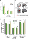
There remains much scientific, clinical, and ethical controversy concerning the use of electroconvulsive therapy (ECT) for psychiatric disorders stemming from a lack of information and knowledge about how such treatment might work, given its nonspecific and spatially unfocused nature. The mode of action of ECT has even been ascribed to a "barbaric" form of placebo effect. Here we show differential, highly specific, spatially distributed effects of ECT on regional brain structure in two populations: patients with unipolar or bipolar disorder. Unipolar and bipolar disorders respond differentially to ECT and the associated local brain-volume changes, which occur in areas previously associated with these diseases, correlate with symptom severity and the therapeutic effect. Our unique evidence shows that electrophysical therapeutic effects, although applied generally, take on regional significance through interactions with brain pathophysiology.
Keywords: hippocampus; magnetic resonance imaging; unipolar depression; voxel-based morphometry.
Conflict of interest statement
The authors declare no conflict of interest.
Figures


References
-
- Weiner RD, Falcone G. Electroconvulsive therapy: How effective is it? J Am Psychiatr Nurses Assoc. 2011;17(3):217–218. - PubMed
-
- Hoy KE, Fitzgerald PB. Brain stimulation in psychiatry and its effects on cognition. Nat Rev Neurol. 2010;6(5):267–275. - PubMed
-
- Wennström M, Hellsten J, Tingström A. Electroconvulsive seizures induce proliferation of NG2-expressing glial cells in adult rat amygdala. Biol Psychiatry. 2004;55(5):464–471. - PubMed
-
- Hellsten J, et al. Electroconvulsive seizures increase hippocampal neurogenesis after chronic corticosterone treatment. Eur J Neurosci. 2002;16(2):283–290. - PubMed
-
- Chen F, Madsen TM, Wegener G, Nyengaard JR. Repeated electroconvulsive seizures increase the total number of synapses in adult male rat hippocampus. Eur Neuropsychopharmacol. 2009;19(5):329–338. - PubMed
Publication types
MeSH terms
LinkOut - more resources
Full Text Sources
Other Literature Sources
Medical

