. 2014 Feb 21;289(8):4882-95.
doi: 10.1074/jbc.M113.501940. Epub 2013 Dec 30.

Retinoblastoma-binding protein 1 has an interdigitated double Tudor domain with DNA binding activity

Affiliations

Retinoblastoma-binding protein 1 has an interdigitated double Tudor domain with DNA binding activity

Weibin Gong et al. J Biol Chem. .

Abstract

Retinoblastoma-binding protein 1 (RBBP1) is a tumor and leukemia suppressor that binds both methylated histone tails and DNA. Our previous studies indicated that RBBP1 possesses a Tudor domain, which cannot bind histone marks. In order to clarify the function of the Tudor domain, the solution structure of the RBBP1 Tudor domain was determined by NMR and is presented here. Although the proteins are unrelated, the RBBP1 Tudor domain forms an interdigitated double Tudor structure similar to the Tudor domain of JMJD2A, which is an epigenetic mark reader. This indicates the functional diversity of Tudor domains. The RBBP1 Tudor domain structure has a significant area of positively charged surface, which reveals a capability of the RBBP1 Tudor domain to bind nucleic acids. NMR titration and isothermal titration calorimetry experiments indicate that the RBBP1 Tudor domain binds both double- and single-stranded DNA with an affinity of 10-100 μM; no apparent DNA sequence specificity was detected. The DNA binding mode and key interaction residues were analyzed in detail based on a model structure of the Tudor domain-dsDNA complex, built by HADDOCK docking using the NMR data. Electrostatic interactions mediate the binding of the Tudor domain with DNA, which is consistent with NMR experiments performed at high salt concentration. The DNA-binding residues are conserved in Tudor domains of the RBBP1 protein family, resulting in conservation of the DNA-binding function in the RBBP1 Tudor domains. Our results provide further insights into the structure and function of RBBP1.

Keywords: DNA-binding Protein; Epigenetics; Nuclear Magnetic Resonance; Protein Structure; Tumor Suppressor Gene.


Figures

FIGURE 1.
FIGURE 1.
Solution structure of the RBBP1 Tudor domain. A and B, ensemble of the top 20 lowest energy structures of the RBBP1 Tudor domain superimposed on HTD-1 (yellow) and HTD-2 (magenta) (A) or on all secondary structure regions (B). C, ribbon representation of the RBBP1 Tudor domain. D, structural alignment of HTD-1 and HTD-2.
FIGURE 2.
FIGURE 2.
Sequence alignment of the human RBBP1 Tudor domain and homologues. A, the alignment of Tudor domains of the RBBP1 family proteins in the whole animal kingdom performed using ClustalW (51) and Espript (52). B, structure-based sequence alignment of Tudor domains of RBBP1 and JMJD2A/2C performed using SSM (53).
FIGURE 3.
FIGURE 3.
Dynamics of the RBBP1 Tudor domain. Shown are steady-state 1H-15N NOE values (A), R1 values (B), R2 values (C), and RMSD values (D) per residue of the RBBP1 Tudor domain. In A–C, mean and mean ± S.D. are shown by dashed lines. The residues from the linker between HTD-1 and HTD-2 are indicated by arrows. The relaxation values of Arg34 in the linker are not shown because the peak of Arg34 is overlapped with other peaks.
FIGURE 4.
FIGURE 4.
Comparison of the RBBP1 and JMJD2A Tudor domain structures. A, overall structural alignment of the RBBP1 Tudor domain (red) and the JMJD2A Tudor domain (green). B, structural alignment of RBBP1 HTD-1/2 (red) and JMJD2A HTD-1/2 (green). C, molecular surface of the RBBP1 Tudor domain (top) and the JMJD2A Tudor domain (bottom) colored according to electrostatic potential. Blue, positively charged; red, negatively charged.
FIGURE 5.
FIGURE 5.
Titration of the RBBP1 Tudor domain with different DNA duplexes. A, 1H-15N HSQC spectrum of the RBBP1 Tudor domain combining together the spectra recorded at various dsDNA2 concentrations. B, chemical shift perturbations for residues Tyr9, Ala18, Glu111, Thr104, and Cys107 as a function of increased dsDNA/protein ratio in the titration experiments. The solid curves are the curve fitting results, from which the KD values were obtained. C, bar diagram of chemical shift perturbations versus residue number at a 1:1 molar ratio of the RBBP1 Tudor domain to different DNA duplexes. D, mapping of CSPs generated by dsDNA2 onto the structure of the RBBP1 Tudor domain. Red, CSP ≥ mean ± S.D.; pink, mean ± S.D. > CSP ≥ mean. E, the electrostatic surface potential of the RBBP1 Tudor domain in the same orientation as D.
FIGURE 6.
FIGURE 6.
Chemical shift perturbation comparisons of various DNA sequences. Shown is a bar diagram of chemical shift perturbations versus residue number at a 1:1 molar ratio of the RBBP1 Tudor domain to different DNA duplexes: dsAT with ssAT (A), dsGC and ssGC (B), dsDNA1 and ssDNA1 (C), and dsDNA2 and ssDNA2 (D).
FIGURE 7.
FIGURE 7.
ITC results of the RBBP1 Tudor domain titrated with DNA. The raw data of heat changes (top panels) and the processed data corresponding to the heat of each injection (bottom panel) are shown for dsAT (A) and ssAT (B).
FIGURE 8.
FIGURE 8.
The structural features which determine the fold of a double interdigitated Tudor domain. A and B, structure-based sequence alignment of RBBP1 HTD-1 with HTD-2 (A) and RBBP1 HTD-1 with the 53BP1 Tudor domain 1 (B). Residues that can be aligned in the structure alignment are capitalized, and residues that cannot be aligned are in lowercase type. Residues within the hydrophobic core are in boldface type and colored red. The N- and C-terminal residues, Ala4 and Asp121, are numbered. C, structure alignment of RBBP1 HTD-1 (dark gray) and the 53BP1 Tudor domain 1 (green). D and E, structure alignment of the hydrophobic core (D) and salt bridges (E) of RBBP1 HTD-1 (magenta) and HTD-2 (yellow).
FIGURE 9.
FIGURE 9.
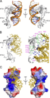
Structural model of the RBBP1 Tudor domain in the complex with dsDNA2. The model was calculated using the HADDOCK Web server (42). A, the best four structures of the top 1 cluster. B, contacts at the interface between the RBBP1 Tudor domain and the DNA duplex in the best structure of cluster 1. The RBBP1 Tudor domain is shown in a ribbon representation, and the residues potentially involved in the binding are shown as sticks. The dsDNA is shown in yellow. C, the electrostatic surface of the RBBP1 Tudor domain in the model of the complex. The charged residues involved in binding are labeled.
FIGURE 10.
FIGURE 10.
Comparison of the binding of DNA to RBBP1, 53BP1, and Esa1 Tudor domains. Ribbon and electrostatic surface representations are shown for the 53BP1 Tudor domain 1 (A), the Esa1 Tudor domain (B), and the RBBP1 Tudor domain (C). DNA binding residues are colored in red in the ribbon representation. DNA binding interfaces are semiopaque in white. Structures in A, B, and the top panel of C are in the same orientation.

References

    1. Malovannaya A., Li Y., Bulynko Y., Jung S. Y., Wang Y., Lanz R. B., O'Malley B. W., Qin J. (2010) Streamlined analysis schema for high-throughput identification of endogenous protein complexes. Proc. Natl. Acad. Sci. U.S.A. 107, 2431–2436 - PMC - PubMed
    1. Wu M. Y., Eldin K. W., Beaudet A. L. (2008) Identification of chromatin remodeling genes Arid4a and Arid4b as leukemia suppressor genes. J. Natl. Cancer Inst. 100, 1247–1259 - PMC - PubMed
    1. Meehan W. J., Samant R. S., Hopper J. E., Carrozza M. J., Shevde L. A., Workman J. L., Eckert K. A., Verderame M. F., Welch D. R. (2004) Breast cancer metastasis suppressor 1 (BRMS1) forms complexes with retinoblastoma-binding protein 1 (RBP1) and the mSin3 histone deacetylase complex and represses transcription. J. Biol. Chem. 279, 1562–1569 - PubMed
    1. Fleischer T. C., Yun U. J., Ayer D. E. (2003) Identification and characterization of three new components of the mSin3A corepressor complex. Mol. Cell Biol. 23, 3456–3467 - PMC - PubMed
    1. Lai A., Kennedy B. K., Barbie D. A., Bertos N. R., Yang X. J., Theberge M. C., Tsai S. C., Seto E., Zhang Y., Kuzmichev A., Lane W. S., Reinberg D., Harlow E., Branton P. E. (2001) RBP1 recruits the mSIN3-histone deacetylase complex to the pocket of retinoblastoma tumor suppressor family proteins found in limited discrete regions of the nucleus at growth arrest. Mol. Cell Biol. 21, 2918–2932 - PMC - PubMed

Publication types

MeSH terms

Associated data

LinkOut - more resources