. 2014 Feb 21;289(8):5228-39.
doi: 10.1074/jbc.M113.510248. Epub 2013 Dec 30.

Mycothiol/mycoredoxin 1-dependent reduction of the peroxiredoxin AhpE from Mycobacterium tuberculosis

Affiliations

Mycothiol/mycoredoxin 1-dependent reduction of the peroxiredoxin AhpE from Mycobacterium tuberculosis

Martín Hugo et al. J Biol Chem. .

Abstract

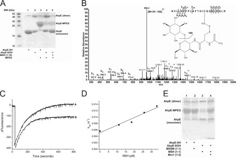
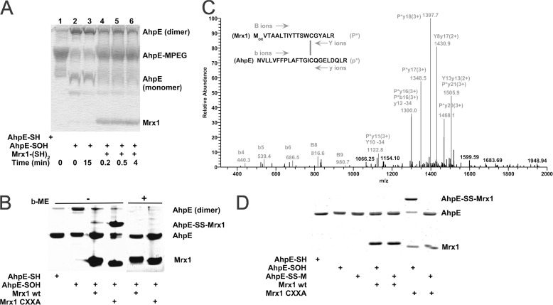
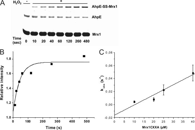
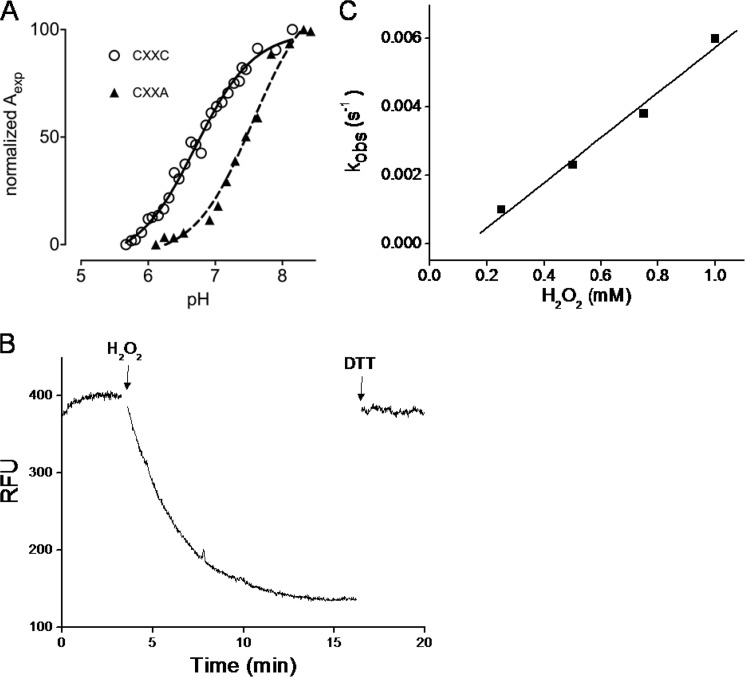
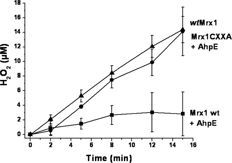
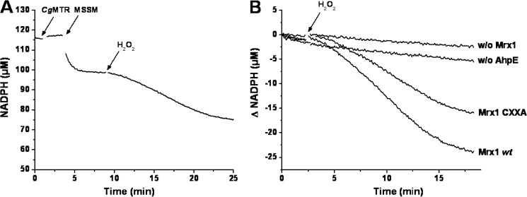
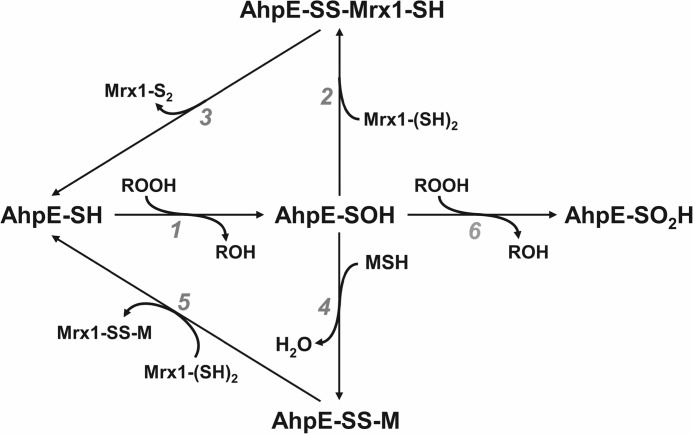
Mycobacterium tuberculosis (M. tuberculosis), the pathogen responsible for tuberculosis, detoxifies cytotoxic peroxides produced by activated macrophages. M. tuberculosis expresses alkyl hydroxyperoxide reductase E (AhpE), among other peroxiredoxins. So far the system that reduces AhpE was not known. We identified M. tuberculosis mycoredoxin-1 (MtMrx1) acting in combination with mycothiol and mycothiol disulfide reductase (MR), as a biologically relevant reducing system for MtAhpE. MtMrx1, a glutaredoxin-like, mycothiol-dependent oxidoreductase, directly reduces the oxidized form of MtAhpE, through a protein mixed disulfide with the N-terminal cysteine of MtMrx1 and the sulfenic acid derivative of the peroxidatic cysteine of MtAhpE. This disulfide is then reduced by the C-terminal cysteine in MtMrx1. Accordingly, MtAhpE catalyzes the oxidation of wt MtMrx1 by hydrogen peroxide but not of MtMrx1 lacking the C-terminal cysteine, confirming a dithiolic mechanism. Alternatively, oxidized MtAhpE forms a mixed disulfide with mycothiol, which in turn is reduced by MtMrx1 using a monothiolic mechanism. We demonstrated the H2O2-dependent NADPH oxidation catalyzed by MtAhpE in the presence of MR, Mrx1, and mycothiol. Disulfide formation involving mycothiol probably competes with the direct reduction by MtMrx1 in aqueous intracellular media, where mycothiol is present at millimolar concentrations. However, MtAhpE was found to be associated with the membrane fraction, and since mycothiol is hydrophilic, direct reduction by MtMrx1 might be favored. The results reported herein allow the rationalization of peroxide detoxification actions inferred for mycothiol, and more recently, for Mrx1 in cellular systems. We report the first molecular link between a thiol-dependent peroxidase and the mycothiol/Mrx1 pathway in Mycobacteria.

Keywords: Hydrogen Peroxide; Mycobacterium tuberculosis; Mycoredoxin; Mycothiol; Peroxiredoxin; Redox Signaling; Thiol.


Figures

FIGURE 1.
FIGURE 1.
Oxidized MtAhpE reacts with MSH forming a mixed disulfide that is reduced by MtMrx1. A, alkylation of MtAhpE with PEG-maleimide. Reduced or oxidized MtAhpE (20 μm) was incubated with or without MSH (200 μm). Proteins were precipitated with TCA, treated with PEG-maleimide (5 mm), and evaluated on a CBB-stained 15% SDS-PAGE. B, identification of S-mycothiolation on cysteine 45 of MtAhpE. Sample was obtained by adding MSH (60 μm) to oxidized MtAhpE (20 μm). The LC-MS/MS spectrum shows data obtained from a 3+ parent ion with m/z = 1026.5. The spectrum displays one major daughter ion at m/z 966.4 corresponding to the neutral loss of inositol (180 Da) after fragmentation at a C-O bond. The y- and b- series of ions allowed exact localization of the mixed disulfide between mycothiol and the cysteine residue. C, kinetics of reaction of oxidized MtAhpE with MSH. Time-dependent decrease (oxidation) and increase (overoxidation) in the intrinsic fluorescence intensity (λex = 295 nm, λem = 340 nm) of MtAhpE (2 μm) in the absence (A) or presence (B) of MSH (18 μm) upon addition of H2O2 (150 μm) in 100 mm sodium phosphate buffer plus 0.1 mm DTPA. D, effect of the MSH concentration on the observed rate constants of MtAhpE intrinsic fluorescence change caused by overoxidation. E, MtAhpE-SS-M reduction by MtMrx1. Reduced (lanes 1 and 2) or oxidized (lanes 3 and 4) MtAhpE (20 μm) was incubated with MSSM (lane 2) or MSH (lanes 3 and 4)(20 μm) for 30 min, followed by incubation without (lane 3) or with MtMrx1 (lane 4) (20 μm) for 15 min. Samples were treated with PEG-maleimide (5 mm) and evaluated by CBB-stained 15% SDS-PAGE.
FIGURE 2.
FIGURE 2.
MtAhpE is reduced by wild type MtMrx1 by a dithiolic mechanism. A, reduced (lane 1) or oxidized (lanes 2–6) MtAhpE (10 μm) incubated in the absence (lanes 1–3) or presence of reduced MtMrx1 (16 μm) for indicated times (lanes 4–6) and treated with 5 mm PEG-maleimide were evaluated on a Coomassie Brilliant Blue (CBB) stained 15% SDS-PAGE. B, reduced and oxidized MtAhpE alone (10 μm), or oxidized MtAhpE incubated with MtMrx1 wt or MtMrx1 CXXA (30 μm) for 15 min were evaluated on a CBB-stained 15% SDS-PAGE in the absence (lanes 1–4, respectively) or presence (lanes 4–8, respectively) of β-ME. A novel band with a molecular mass compatible with a mixed disulfide formation between MtAhpE and MtMrx1CXXA is indicated as MtAhpE-SS-MtMrx1. C, mass spectrometric analysis of the MtAhpE-SS-MtMrx1 complex is shown in Fig. 2B. A quadruply charged parent ion of [M+4H]4+ = 1183.7 Da shows fragmentation characteristics of a disulfide linkage between Cys17 of MtMrx1 and Cys45 of MtAhpE, as determined by the DBond software (29). P*, one strand of a dipeptide; p*, the other strand of a dipeptide; capital letters, fragment ions from peptide P*; lowercase letters, fragment ions from peptide p*. The loss of 34 atomic mass units represents formation of dehydroalanine from C-S bond fragmentation. D, reaction of MtAhpE-SS-M with MtMrx1CXXA does not form a protein-protein intermolecular mixed disulfide. Reduced (lane 1) and oxidized MtAhpE (lanes 2–7) alone (lanes 2, 4, and 6), or incubated with MSH during 30 min (lanes 3, 5, and 7), were incubated in the absence (lanes 2 and 3) or presence of MtMrx1 wt (lanes 4 and 5) or MtMrx1CXXA (lanes 6 and 7) for 15 min. Reaction was stopped by addition of 5 mm NEM, and samples were evaluated on a CBB-stained 15% SDS-PAGE under non-reducing conditions. A novel band with a molecular mass compatible with a mixed disulfide formation between MtAhpE and MtMrx1CXXA is indicated as MtAhpE-SS-MtMrx1.
FIGURE 3.
FIGURE 3.
Kinetics of the reaction of MtAhpE-SOH with the nucleophilic thiol in MtMrx1CXXA. A, oxidized MtAhpE (1 μm) was incubated with MtMrx1CXXA (25 μm), aliquots were taken at different incubation times and the reaction was stopped by addition of 10% TCA. Samples were evaluated on a CBB-stained 15% SDS-PAGE. B, time-dependent increase of the relative band intensity of the mixed disulfide shown in A, expressed as MtAhpE-SS-MtMrx1/MtMrx1CXXA. MtMrx1CXXA in concentrations of more than 10 times excess remain constant, and was used as protein load control. The continuous line shows the best fit to an exponential curve. C, effect of increasing MtMrx1CXXA concentrations on the observed rate constants of intermolecular disulfide formation.
FIGURE 4.
FIGURE 4.
MtMrx1 thiol pKa determinations and kinetics of oxidation by H2O2. A, pKa titration curves for wild type (circles) (15) and the CXXA mutant (triangles). The specific absorption of the thiolate ion at 240 nm as a function of the pH is shown. Aexp is determined as described (31). Data were fitted with the Henderson-Hasselbach equation. B, time-dependent decrease in the total intrinsic fluorescence intensity (λex = 295 nm, λem = 335 nm) of MtMrx1wt (10 μm) upon oxidation by H2O2 in 100 mm sodium phosphate buffer plus 0.1 mm DTPA, at pH 7.4 and room temperature. The first arrow indicates the addition of excess H2O2 (1.5 mm) and the second, the addition of DTT (1.5 mm). C, effect of H2O2 concentration on the observed rate constants of wtMtMrx1 intrinsic fluorescence change.
FIGURE 5.
FIGURE 5.
MtAhpE catalyzes H2O2 reduction in the presence of MtMrx1. H2O2 was infused (J = 1 μm min−1) to reaction mixtures containing 50 μm reduced MtMrx1 wt (triangles), 50 μm reduced MtMrx1 CXXA + 2 μm MtAhpE (circles), or 50 μm reduced MtMrx1 wt + 2 μm MtAhpE (squares). Remaining H2O2 was measured at different time points, using Amplex®red oxidation assay. Data points represent an average of n = 3 ± S.D.
FIGURE 6.
FIGURE 6.
NADPH consumption during MtAhpE-mediated H2O2 reduction. A, time-dependent consumption of NADPH (100 μm) in a coupled assay containing 20 μm MtAhpE, 5 μm MtMrx1wt, 0.13 μm CgMR, 30 μm MSSM, and 20 μm H2O2 in 50 mm HEPES, 0.5 mm EDTA, pH 7.8 at 25 °C. The arrows indicate the addition of the last three mentioned components to the mixture. B, NADPH consumption upon addition of H2O2 (arrow) in mixtures as in A (Mrx1 wt); with CXXA instead of wt MtMrx1 (Mrx1 CXXA); in the absence of MtMrx1 (w/o Mrx1) or in the absence of MtAhpE (w/o AhpE). Representative traces that were repeated in independent days with the same results are shown.
FIGURE 7.
FIGURE 7.
Mechanisms proposed for MSH/Mrx1-dependent MtAhpE reduction. MtAhpE is oxidized by the peroxide to form a sulfenic acid (reaction 1). Sulfenic acid is then directly reduced by MtMrx1 (reactions 2 and 3), or through an intermediate disulfide formation with mycothiol (reaction 4), followed by reduction by MtMrx1 (reaction 5). The leaving Mrx1-S2 and Mrx1-SS-M disulfide species are then reduced by a second mycothiol molecule forming mycothiol disulfide (MSSM) and reduced MtMrx1 as reported (7, 15). The formed MSSM is in turn reduced by the NADPH dependent flavoenzyme, mycothiol disulfide reductase (MR), as previously reported (8, 15). Both reducing pathways may compete with enzyme oxidative inactivation (overoxidation) to a sulfinic acid (reaction 6). The lower pathway (reactions 4–5) would predominate in the cytosol while the upper one (reactions 2–3) could be favored in membrane-associated compartments due to the hydrophilic nature of MSH.

Similar articles

Cited by

  • Relevance of peroxiredoxins in pathogenic microorganisms.
    de Oliveira MA, Tairum CA, Netto LES, de Oliveira ALP, Aleixo-Silva RL, Cabrera VIM, Breyer CA, Dos Santos MC. de Oliveira MA, et al. Appl Microbiol Biotechnol. 2021 Aug;105(14-15):5701-5717. doi: 10.1007/s00253-021-11360-5. Epub 2021 Jul 14. Appl Microbiol Biotechnol. 2021. PMID: 34258640 Review.
  • European contribution to the study of ROS: A summary of the findings and prospects for the future from the COST action BM1203 (EU-ROS).
    Egea J, Fabregat I, Frapart YM, Ghezzi P, Görlach A, Kietzmann T, Kubaichuk K, Knaus UG, Lopez MG, Olaso-Gonzalez G, Petry A, Schulz R, Vina J, Winyard P, Abbas K, Ademowo OS, Afonso CB, Andreadou I, Antelmann H, Antunes F, Aslan M, Bachschmid MM, Barbosa RM, Belousov V, Berndt C, Bernlohr D, Bertrán E, Bindoli A, Bottari SP, Brito PM, Carrara G, Casas AI, Chatzi A, Chondrogianni N, Conrad M, Cooke MS, Costa JG, Cuadrado A, My-Chan Dang P, De Smet B, Debelec-Butuner B, Dias IHK, Dunn JD, Edson AJ, El Assar M, El-Benna J, Ferdinandy P, Fernandes AS, Fladmark KE, Förstermann U, Giniatullin R, Giricz Z, Görbe A, Griffiths H, Hampl V, Hanf A, Herget J, Hernansanz-Agustín P, Hillion M, Huang J, Ilikay S, Jansen-Dürr P, Jaquet V, Joles JA, Kalyanaraman B, Kaminskyy D, Karbaschi M, Kleanthous M, Klotz LO, Korac B, Korkmaz KS, Koziel R, Kračun D, Krause KH, Křen V, Krieg T, Laranjinha J, Lazou A, Li H, Martínez-Ruiz A, Matsui R, McBean GJ, Meredith SP, Messens J, Miguel V, Mikhed Y, Milisav I, Milković L, Miranda-Vizuete A, Mojović M, Monsalve M, Mouthuy PA, Mulvey J, Münzel T, Muzykantov V, Nguyen ITN, Oelze M, Oliveira NG, Palmeira CM, Papaevgeniou N, Pavićević A, Pedre B, Peyrot F, Phy… See abstract for full author list ➔ Egea J, et al. Redox Biol. 2017 Oct;13:94-162. doi: 10.1016/j.redox.2017.05.007. Epub 2017 May 18. Redox Biol. 2017. PMID: 28577489 Free PMC article. Review.
  • Peroxiredoxins wear many hats: Factors that fashion their peroxide sensing personalities.
    Bolduc J, Koruza K, Luo T, Malo Pueyo J, Vo TN, Ezeriņa D, Messens J. Bolduc J, et al. Redox Biol. 2021 Jun;42:101959. doi: 10.1016/j.redox.2021.101959. Epub 2021 Apr 20. Redox Biol. 2021. PMID: 33895094 Free PMC article. Review.
  • Reactive species and pathogen antioxidant networks during phagocytosis.
    Piacenza L, Trujillo M, Radi R. Piacenza L, et al. J Exp Med. 2019 Mar 4;216(3):501-516. doi: 10.1084/jem.20181886. Epub 2019 Feb 21. J Exp Med. 2019. PMID: 30792185 Free PMC article. Review.
  • Oxidative Stress-Generating Antimicrobials, a Novel Strategy to Overcome Antibacterial Resistance.
    Mourenza Á, Gil JA, Mateos LM, Letek M. Mourenza Á, et al. Antioxidants (Basel). 2020 Apr 26;9(5):361. doi: 10.3390/antiox9050361. Antioxidants (Basel). 2020. PMID: 32357394 Free PMC article. Review.

References

    1. World Health Organization (2012) Global Tuberculosis Report
    1. Nathan C. (2009) Taming tuberculosis: a challenge for science and society. Cell Host Microbe. 5, 220–224 - PubMed
    1. Alvarez M. N., Peluffo G., Piacenza L., Radi R. (2011) Intraphagosomal peroxynitrite as a macrophage-derived cytotoxin against internalized Trypanosoma cruzi: consequences for oxidative killing and role of microbial peroxiredoxins in infectivity. J. Biol. Chem. 286, 6627–6640 - PMC - PubMed
    1. Fang F. C. (2004) Antimicrobial reactive oxygen and nitrogen species: concepts and controversies. Nat. Rev. Microbiol. 2, 820–832 - PubMed
    1. Nathan C., Shiloh M. U. (2000) Reactive oxygen and nitrogen intermediates in the relationship between mammalian hosts and microbial pathogens. Proc. Natl. Acad. Sci. U.S.A. 97, 8841–8848 - PMC - PubMed

Publication types

LinkOut - more resources