Bone morphogenetic protein-7 accelerates fracture healing in osteoporotic rats
- PMID: 24379457
- PMCID: PMC3868133
- DOI: 10.4103/0019-5413.121569
Bone morphogenetic protein-7 accelerates fracture healing in osteoporotic rats
Abstract
Background: Osteoporosis is characterized by low bone mass, bone fragility and increased susceptibility to fracture. Fracture healing in osteoporosis is delayed and rates of implant failure are high with few biological treatment options available. This study aimed to determine whether a single dose of bone morphogenetic protein-7 (BMP-7) in a collagen/carboxy-methyl cellulose (CMC) composite enhanced fracture healing in an osteoporotic rat model.
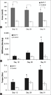
Materials and methods: An open femoral midshaft osteotomy was performed in female rats 3 months post-ovarectomy. Rats were randomized to receive either BMP-7 composite (n = 30) or composite alone (n = 30) at the fracture site during surgery. Thereafter calluses were collected on days 12, 20 and 31. Callus cross-sectional area, bone mineral density, biomechanical stiffness and maximum torque, radiographic bony union and histological callus maturity were evaluated at each time point.
Results: There were statistically significant increases in bone mineral density and callus cross-section area at all time points in the BMP-7 group as compared to controls and biomechanical readings showed stronger bones at day 31 in the BMP-7 group. Histological and radiographic evaluation indicated significant acceleration of bony union in the BMP-7 group as compared to controls.
Conclusion: This study demonstrated that BMP-7 accelerates fracture healing in an oestrogen-deficient environment in a rat femoral fracture healing model to scientific relevance level I. The use of BMP-7 composite could offer orthopedic surgeons an advantage over oestrogen therapy, enhancing osteoporotic fracture healing with a single, locally applied dose at the time of surgery, potentially overcoming delays in healing caused by the osteoporotic state.
Keywords: Bone morphogenetic protein-7; carboxy-methyl cellulose; oestrogen; osteoporotic fracture healing; rat femur.
Conflict of interest statement
Figures


Similar articles
-
Do osteoporotic fractures constitute a greater recalcitrant challenge for skeletal regeneration? Investigating the efficacy of BMP-7 and zoledronate treatment of diaphyseal fractures in an open fracture osteoporotic rat model.Osteoporos Int. 2017 Feb;28(2):697-707. doi: 10.1007/s00198-016-3771-8. Epub 2016 Nov 7. Osteoporos Int. 2017. PMID: 27822590 Free PMC article.
-
Direct adenoviral transfer of bone morphogenetic protein-2 cDNA enhances fracture healing in osteoporotic sheep.Hum Gene Ther. 2006 May;17(5):507-17. doi: 10.1089/hum.2006.17.507. Hum Gene Ther. 2006. PMID: 16716108
-
Augmentation of fracture healing by hydroxyapatite/collagen paste and bone morphogenetic protein-2 evaluated using a rat femur osteotomy model.J Orthop Res. 2018 Jan;36(1):129-137. doi: 10.1002/jor.23646. Epub 2017 Jul 31. J Orthop Res. 2018. PMID: 28681967
-
Effect of osteoporosis medications on fracture healing.Osteoporos Int. 2016 Mar;27(3):861-871. doi: 10.1007/s00198-015-3331-7. Epub 2015 Sep 29. Osteoporos Int. 2016. PMID: 26419471 Review.
-
Fracture healing in osteoporotic bone.Injury. 2016 Jun;47 Suppl 2:S21-6. doi: 10.1016/S0020-1383(16)47004-X. Injury. 2016. PMID: 27338222 Review.
Cited by
-
Arsenic Induces Differential Neurotoxicity in Male, Female, and E2-Deficient Females: Comparative Effects on Hippocampal Neurons and Cognition in Adult Rats.Mol Neurobiol. 2022 May;59(5):2729-2744. doi: 10.1007/s12035-022-02770-1. Epub 2022 Feb 17. Mol Neurobiol. 2022. PMID: 35175559
-
Bone morphogenic proteins are a good choice for select spinal surgeries and merit further research.J Spine Surg. 2017 Mar;3(1):119-121. doi: 10.21037/jss.2017.02.03. J Spine Surg. 2017. PMID: 28435931 Free PMC article. No abstract available.
-
Recombinant Human Peptide Growth Factors, Bone Morphogenetic Protein-7 (rhBMP7), and Platelet-Derived Growth Factor-BB (rhPDGF-BB) for Osteoporosis Treatment in an Oophorectomized Rat Model.Biomolecules. 2024 Mar 7;14(3):317. doi: 10.3390/biom14030317. Biomolecules. 2024. PMID: 38540737 Free PMC article.
-
Biomechanical Characteristics of Osteoporotic Fracture Healing in Ovariectomized Rats: A Systematic Review.PLoS One. 2016 Apr 7;11(4):e0153120. doi: 10.1371/journal.pone.0153120. eCollection 2016. PLoS One. 2016. PMID: 27055104 Free PMC article.
-
Analysis of fracture healing in osteopenic bone caused by disuse: experimental study.Braz J Med Biol Res. 2016 Mar;49(3):e5076. doi: 10.1590/1414-431X20155076. Epub 2016 Feb 2. Braz J Med Biol Res. 2016. PMID: 26840708 Free PMC article.
References
-
- Walsh WR, Sherman P, Howlett CR, Sonnabend DH, Ehrlich MG. Fracture healing in a rat osteopenia model. Clin Orthop Relat Res. 1997:218–27. - PubMed
-
- Moazzaz P, Gupta MC, Gilotra MM, Gilotra MN, Maitra S, Theerajunyaporn T, et al. Estrogen-dependent actions of bone morphogenetic protein-7 on spine fusion in rats. Spine (Phila Pa 1976) 2005;30:1706–11. - PubMed
-
- Namkung-Matthai H, Appleyard R, Jansen J, Hao Lin J, Maastricht S, Swain M, et al. Osteoporosis influences the early period of fracture healing in a rat osteoporotic model. Bone. 2001;28:80–6. - PubMed
-
- McLain RF, McKinley TO, Yerby SA, Smith TS, Sarigul-Klijn N. The effect of bone quality on pedicle screw loading in axial instability. A synthetic model. Spine (Phila Pa 1976) 1997;22:1454–60. - PubMed
-
- Halvorson TL, Kelley LA, Thomas KA, Whitecloud TS, 3rd, Cook SD. Effects of bone mineral density on pedicle screw fixation. Spine (Phila Pa 1976) 1994;19:2415–20. - PubMed
LinkOut - more resources
Full Text Sources
Other Literature Sources
