Review of cystic and solid tumors of the iris
- PMID: 24379549
- PMCID: PMC3872564
- DOI: 10.4103/0974-620X.122269
Review of cystic and solid tumors of the iris
Abstract
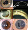
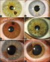
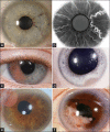
Iris tumors are broadly classified into cystic or solid lesions. The cystic lesions arise from iris pigment epithelium (IPE) or iris stroma. IPE cysts classically remain stable without need for intervention. Iris stromal cyst, especially those in newborns, usually requires therapy of aspiration, possibly with alcohol-induced sclerosis, or surgical resection. The solid tumors included melanocytic and nonmelanocytic lesions. The melanocytic iris tumors include freckle, nevus (including melanocytoma), Lisch nodule, and melanoma. Information from a tertiary referral center revealed that transformation of suspicious iris nevus to melanoma occurred in 4% by 10 years and 11% by 20 years. Risk factors for transformation of iris nevus to melanoma can be remembered using the ABCDEF guide as follows: A=age young (<40 years), B=blood (hyphema) in anterior chamber, C=clock hour of mass inferiorly, D=diffuse configuration, E=ectropion, F=feathery margins. The most powerful factors are diffuse growth pattern and hyphema. Tumor seeding into the anterior chamber angle and onto the iris stroma are also important. The nonmelanocytic iris tumors are relatively uncommon and included categories of choristomatous, vascular, fibrous, neural, myogenic, epithelial, xanthomatous, metastatic, lymphoid, leukemic, secondary, and non-neoplastic simulators. Overall, the most common diagnoses in a clinical series include nevus, IPE cyst, and melanoma. In summary, iris tumors comprise a wide spectrum including mostly iris nevus, IPE cyst, and iris melanoma. Risk factors estimating transformation of iris nevus to melanoma can be remembered by the ABCDEF guide.
Keywords: Cyst; eye; iris; melanoma; metastasis; nevus; tumor.
Conflict of interest statement
Figures


References
-
- Shields CL, Kancherla S, Patel J, Vijayvargiya P, Suriano MM, Kolbus E, et al. Clinical survey of 3680 iris tumors based on patient age at presentation. Ophthalmology. 2012;119:407–14. - PubMed
-
- Shields JA, Shields CL. An Atlas and Textbook. 2nd ed. Philadelphia: Lippincott Williams and Wilkins; 2008. Intraocular Tumors; pp. 3–58.
-
- Duke JR, Dunn SN. Primary tumors of the iris. Arch Ophthalmol. 1958;59:204–14. - PubMed
Publication types
LinkOut - more resources
Full Text Sources
Other Literature Sources

