

Assessment of the diagnostic performance and interobserver variability of endocytoscopy in Barrett's esophagus: a pilot ex-vivo study
- PMID: 24379583
- PMCID: PMC3870511
- DOI: 10.3748/wjg.v19.i46.8652
Assessment of the diagnostic performance and interobserver variability of endocytoscopy in Barrett's esophagus: a pilot ex-vivo study
Abstract
Aim: To investigate a classification of endocytoscopy (ECS) images in Barrett's esophagus (BE) and evaluate its diagnostic performance and interobserver variability.
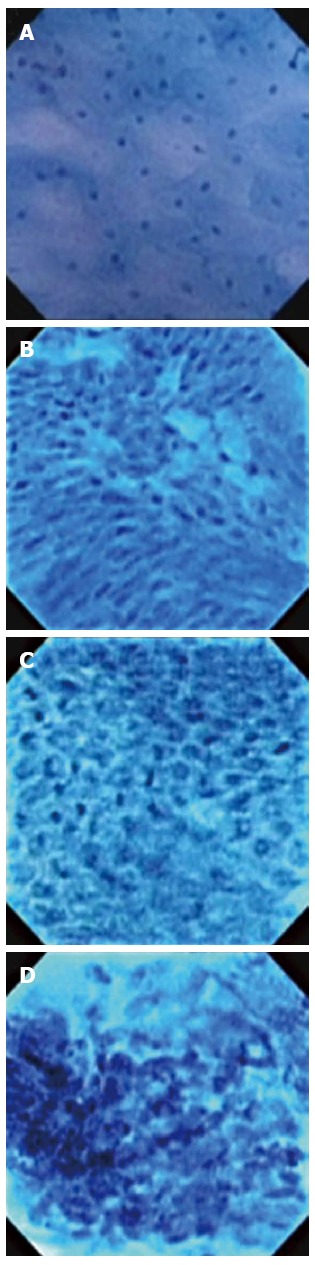
Methods: ECS was applied to surveillance endoscopic mucosal resection (EMR) specimens of BE ex-vivo. The mucosal surface of specimen was stained with 1% methylene blue and surveyed with a catheter-type endocytoscope. We selected still images that were most representative of the endoscopically suspect lesion and matched with the final histopathological diagnosis to accomplish accurate correlation. The diagnostic performance and inter-observer variability of the new classification scheme were assessed in a blinded fashion by physicians with expertise in both BE and ECS and inexperienced physicians with no prior exposure to ECS.
Results: Three staff physicians and 22 gastroenterology fellows classified eight randomly assigned unknown still ECS pictures (two images per each classification) into one of four histopathologic categories as follows: (1) BEC1-squamous epithelium; (2) BEC2-BE without dysplasia; (3) BEC3-BE with dysplasia; and (4) BEC4-esophageal adenocarcinoma (EAC) in BE. Accuracy of diagnosis in staff physicians and clinical fellows were, respectively, 100% and 99.4% for BEC1, 95.8% and 83.0% for BEC2, 91.7% and 83.0% for BEC3, and 95.8% and 98.3% for BEC4. Interobserver agreement of the faculty physicians and fellows in classifying each category were 0.932 and 0.897, respectively.
Conclusion: This is the first study to investigate classification system of ECS in BE. This ex-vivo pilot study demonstrated acceptable diagnostic accuracy and excellent interobserver agreement.
Keywords: Barrett’s esophagus; Dysplasia; Endocytoscopy; Esophageal adenocarcinoma; Interobserver agreement.
Figures
Similar articles
-
Development and Validation of a Classification System to Identify High-Grade Dysplasia and Esophageal Adenocarcinoma in Barrett's Esophagus Using Narrow-Band Imaging.Gastroenterology. 2016 Mar;150(3):591-8. doi: 10.1053/j.gastro.2015.11.037. Epub 2015 Nov 25. Gastroenterology. 2016. PMID: 26627609
-
Observer agreement in the assessment of narrowband imaging system surface patterns in Barrett's esophagus: a multicenter study.Endoscopy. 2011 Sep;43(9):745-51. doi: 10.1055/s-0030-1256631. Epub 2011 Aug 10. Endoscopy. 2011. PMID: 21833901
-
Endoscopic mucosal resection results in change of histologic diagnosis in Barrett's esophagus patients with visible and flat neoplasia: a multicenter cohort study.Dig Dis Sci. 2013 Jun;58(6):1703-9. doi: 10.1007/s10620-013-2689-7. Epub 2013 Apr 30. Dig Dis Sci. 2013. PMID: 23633158 Free PMC article. Clinical Trial.
-
Barrett's oesophagus: the new endoscopic modalities have a future.Gut. 2005 Mar;54 Suppl 1(Suppl 1):i33-7. doi: 10.1136/gut.2004.041574. Gut. 2005. PMID: 15711006 Free PMC article. Review.
-
Total endoscopic eradication of Barrett's esophagus: study methodology, candidate selection, and clinical outcomes.Endoscopy. 2008 Dec;40(12):994-9. doi: 10.1055/s-2008-1077785. Epub 2008 Dec 8. Endoscopy. 2008. PMID: 19065482 Review. No abstract available.
Cited by
-
Future of Endoscopy in Inflammatory Bowel Diseases (IBDs).Cureus. 2022 Sep 25;14(9):e29567. doi: 10.7759/cureus.29567. eCollection 2022 Sep. Cureus. 2022. PMID: 36312686 Free PMC article. Review.
-
Clinical Efficacy of Endocytoscopy for Gastrointestinal Endoscopy.Clin Endosc. 2021 Jul;54(4):455-463. doi: 10.5946/ce.2021.165. Epub 2021 Jul 7. Clin Endosc. 2021. PMID: 34233111 Free PMC article.
-
Optical Biopsy of Dysplasia in Barrett's Oesophagus Assisted by Artificial Intelligence.Cancers (Basel). 2023 Mar 24;15(7):1950. doi: 10.3390/cancers15071950. Cancers (Basel). 2023. PMID: 37046611 Free PMC article.
-
Advances in optical gastrointestinal endoscopy: a technical review.Mol Oncol. 2021 Oct;15(10):2580-2599. doi: 10.1002/1878-0261.12792. Epub 2020 Sep 19. Mol Oncol. 2021. PMID: 32915503 Free PMC article. Review.
-
Endoscopic imaging of Barrett's esophagus.World J Gastrointest Endosc. 2016 Mar 10;8(5):259-66. doi: 10.4253/wjge.v8.i5.259. World J Gastrointest Endosc. 2016. PMID: 26981177 Free PMC article. Review.
References
-
- Gorospe EC, Leggett CL, Sun G, Anderson MA, Gupta M, Penfield JD, Lutzke L, Lewis JT, Wong Kee Song LM, Wang KK. Diagnostic performance of two confocal endomicroscopy systems in detecting Barrett’s dysplasia: a pilot study using a novel bioprobe in ex vivo tissue. Gastrointest Endosc. 2012;76:933–938. - PubMed
-
- Wallace MB, Crook JE, Saunders M, Lovat L, Coron E, Waxman I, Sharma P, Hwang JH, Banks M, DePreville M, et al. Multicenter, randomized, controlled trial of confocal laser endomicroscopy assessment of residual metaplasia after mucosal ablation or resection of GI neoplasia in Barrett’s esophagus. Gastrointest Endosc. 2012;76:539–47.e1. - PubMed
-
- Wolfsen HC, Crook JE, Krishna M, Achem SR, Devault KR, Bouras EP, Loeb DS, Stark ME, Woodward TA, Hemminger LL, et al. Prospective, controlled tandem endoscopy study of narrow band imaging for dysplasia detection in Barrett’s Esophagus. Gastroenterology. 2008;135:24–31. - PubMed
-
- Sharma P, Hawes RH, Bansal A, Gupta N, Curvers W, Rastogi A, Singh M, Hall M, Mathur SC, Wani SB, et al. Standard endoscopy with random biopsies versus narrow band imaging targeted biopsies in Barrett’s oesophagus: a prospective, international, randomised controlled trial. Gut. 2013;62:15–21. - PubMed
Publication types
MeSH terms
Supplementary concepts
LinkOut - more resources
Full Text Sources
Other Literature Sources
Medical
