

Measurement of retinal nerve fiber layer thickness in eyes with optic disc swelling by using scanning laser polarimetry and optical coherence tomography
- PMID: 24379653
- PMCID: PMC3872170
- DOI: 10.2147/OPTH.S46769
Measurement of retinal nerve fiber layer thickness in eyes with optic disc swelling by using scanning laser polarimetry and optical coherence tomography
Abstract
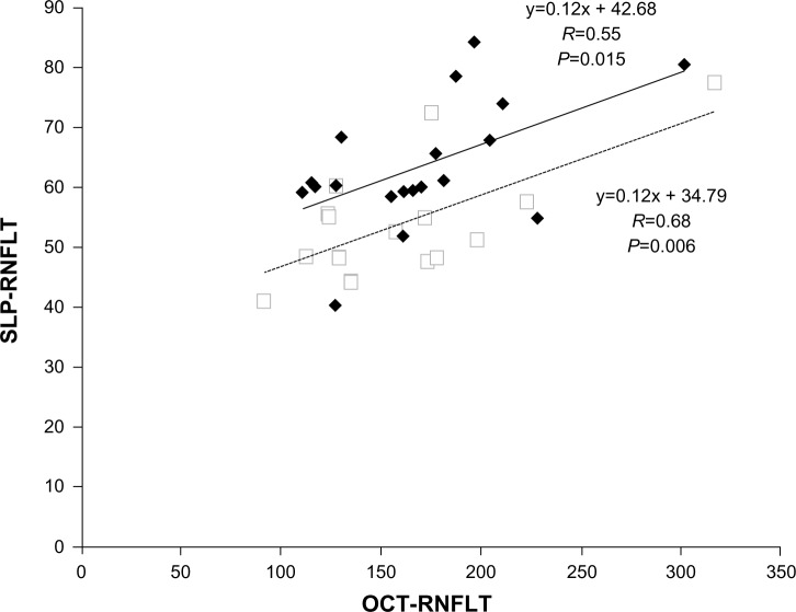
Background: The retinal nerve fiber layer thickness (RNFLT) in patients with optic disc swelling of different etiologies was compared using scanning laser polarimetry (SLP) and spectral-domain optical coherence tomography (OCT).
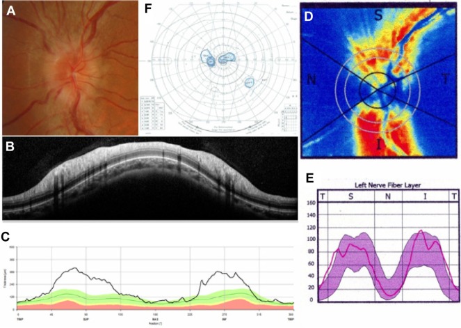
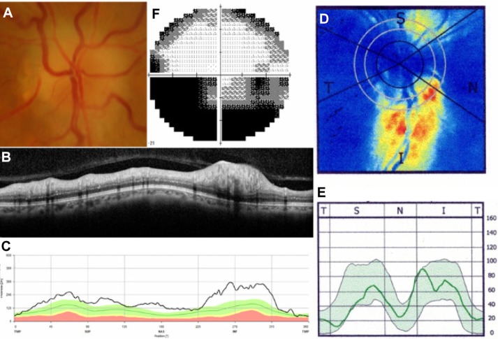
Methods: Forty-seven patients with optic disc swelling participated in the cross-sectional study. Both GDx SLP (enhanced corneal compensation) and Spectralis spectral-domain OCT measurements of RNFLT were made in 19 eyes with papilledema (PE), ten eyes with optic neuritis (ON), and 18 eyes with nonarteritic anterior ischemic optic neuropathy (NAION) at the neuro-ophthalmology clinic at Kyoto University Hospital. Differences in SLP (SLP-RNFLT) and OCT (OCT-RNFLT) measurements among different etiologies were investigated.
Results: No statistical differences in average OCT-RNFLT among PE, ON, and NAION patients were noted. Average SLP-RNFLT in NAION patients was smaller than in PE (P<0.01) or ON (P=0.02) patients. When RNFLT in each retinal quadrant was compared, no difference among etiologies was noted on OCT, but on SLP, the superior quadrant was thinner in NAION than in PE (P<0.001) or ON (P=0.001) patients. Compared with age-adjusted normative data of SLP-RNFLT, average SLP-RNFLT in PE (P<0.01) and ON (P<0.01) patients was greater. Superior SLP-RNFLT in NAION patients was smaller (P=0.026). The ratio of average SLP-RNFLT to average OCT-RNFLT was smaller in NAION than in PE (P=0.001) patients.
Conclusion: In the setting of RNFL thickening, despite increased light retardance in PE and ON eyes, SLP revealed that NAION eyes have less retardance, possibly associated with ischemic axonal loss.
Keywords: optic disc swelling; optical coherence tomography; scanning laser polarimetry.
Figures
Similar articles
-
Scanning laser polarimetry reveals status of RNFL integrity in eyes with optic nerve head swelling by OCT.Invest Ophthalmol Vis Sci. 2012 Apr 18;53(4):1962-70. doi: 10.1167/iovs.11-9339. Invest Ophthalmol Vis Sci. 2012. PMID: 22410562 Free PMC article.
-
Scanning laser polarimetry, but not optical coherence tomography predicts permanent visual field loss in acute nonarteritic anterior ischemic optic neuropathy.Invest Ophthalmol Vis Sci. 2013 Aug 15;54(8):5514-9. doi: 10.1167/iovs.13-12253. Invest Ophthalmol Vis Sci. 2013. PMID: 23838768 Free PMC article.
-
Quantitative assessment of optic nerve head morphology and retinal nerve fibre layer in non-arteritic anterior ischaemic optic neuropathy with optical coherence tomography and confocal scanning laser ophthalmoloscopy.Br J Ophthalmol. 2009 Jun;93(6):731-5. doi: 10.1136/bjo.2008.143297. Epub 2009 Feb 11. Br J Ophthalmol. 2009. PMID: 19211599
-
[Nonarteritic ischemic optic neuropathy animal model and its treatment applications].Nippon Ganka Gakkai Zasshi. 2014 Apr;118(4):331-61. Nippon Ganka Gakkai Zasshi. 2014. PMID: 24864434 Review. Japanese.
-
[New insights into the study of optic nerve diseases].Nippon Ganka Gakkai Zasshi. 2013 Mar;117(3):187-210; discussion 211. Nippon Ganka Gakkai Zasshi. 2013. PMID: 23631254 Review. Japanese.
Cited by
-
Ophthalmic Manifestations of Hematopoietic Malignancy.Case Rep Ophthalmol Med. 2016;2016:6074968. doi: 10.1155/2016/6074968. Epub 2016 Jun 7. Case Rep Ophthalmol Med. 2016. PMID: 27375913 Free PMC article.
-
Optical coherence tomography angiography of optic disc perfusion in non-arteritic anterior ischemic optic neuropathy.Int J Ophthalmol. 2017 Sep 18;10(9):1402-1406. doi: 10.18240/ijo.2017.09.12. eCollection 2017. Int J Ophthalmol. 2017. PMID: 28944200 Free PMC article.
References
-
- Jonas JB, Dichtl A. Evaluation of the retinal nerve fiber layer. Surv Ophthalmol. 1996;40(5):369–378. - PubMed
-
- Pasol J. Neuro-ophthalmic disease and optical coherence tomography: glaucoma look-alikes. Curr Opin Ophthalmol. 2011;22(2):124–132. - PubMed
-
- Burk RO, Völcker HE. Current imaging of the optic disk and retinal nerve fiber layer. Curr Opin Ophthalmol. 1996;7(2):99–108. - PubMed
LinkOut - more resources
Full Text Sources
Other Literature Sources
Miscellaneous
