

Glucocorticoid programing of the mesopontine cholinergic system
- PMID: 24379803
- PMCID: PMC3862116
- DOI: 10.3389/fendo.2013.00190
Glucocorticoid programing of the mesopontine cholinergic system
Abstract
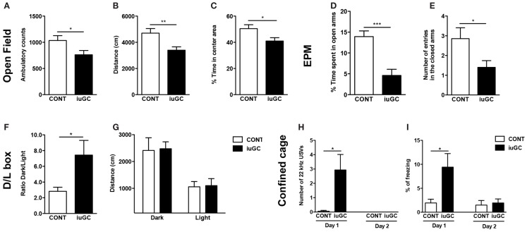
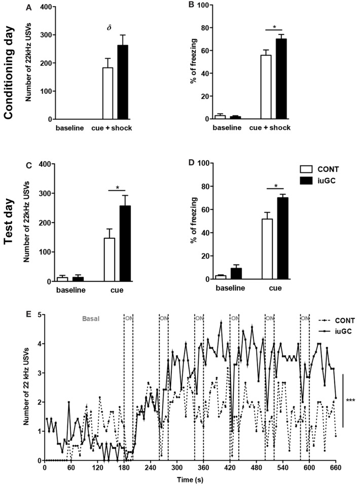
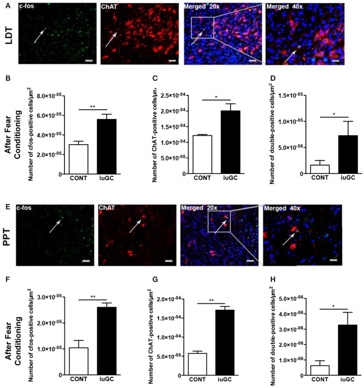
Stress perception, response, adaptation, and coping strategies are individually distinct, and the sequel of stress and/or glucocorticoids (GCs) is also distinct between subjects. In the last years, it has become clear that early life stress is a powerful modulator of neuroendocrine stress-responsive circuits, programing intrinsic susceptibility to stress, and potentiating the appearance of stress-related disorders such as depression, anxiety, and addiction. Herein we were interested in understanding how early life experiences reset the normal processing of negative stimuli, leading to emotional dysfunction. Animals prenatally exposed to GCs (in utero glucocorticoid exposure, iuGC) present hyperanxiety, increased fear behavior, and hyper-reactivity to negative stimuli. In parallel, we found a remarkable increase in the number of aversive 22 kHz ultrasonic vocalizations in response to an aversive cue. Considering the suggested role of the mesopontine tegmentum cholinergic pathway, arising from the laterodorsal tegmental nucleus (LDT) and pedunculopontine tegmental nucleus (PPT), in the initiation of 22 kHz vocalizations and hypothetically in the control of emotional arousal and tone, we decided to evaluate the condition of this circuit in iuGC animals. Notably, in a basal situation, iuGC animals present increased choline acetyltransferase (ChAT) expression in the LDT and PPT, but not in other cholinergic nuclei, namely in the nucleus basalis of Meynert. In addition, and in accordance with the amplified response to an adverse stimulus of iuGC animals, we found marked changes in the cholinergic activation pattern of LDT and PPT regions. Altogether, our results suggest a specific cholinergic pathway programing by prenatal GC, and hint that this may be of relevance in setting individual stress vulnerability threshold.
Keywords: acetylcholine; anxiety; fear; glucocorticoids; laterodorsal tegmental nucleus; pedunculopontine tegmental nucleus; stress; ultrasonic vocalizations.
Figures


Similar articles
-
Ascending projections from the pedunculopontine tegmental nucleus and the adjacent mesopontine tegmentum in the rat.J Comp Neurol. 1988 Aug 22;274(4):483-515. doi: 10.1002/cne.902740403. J Comp Neurol. 1988. PMID: 2464621
-
Cholinergic and non-cholinergic projections from the pedunculopontine and laterodorsal tegmental nuclei to the medial geniculate body in Guinea pigs.Front Neuroanat. 2010 Oct 19;4:137. doi: 10.3389/fnana.2010.00137. eCollection 2010. Front Neuroanat. 2010. PMID: 21060717 Free PMC article.
-
Selective decrease of cholinergic signaling from pedunculopontine and laterodorsal tegmental nuclei has little impact on cognition but markedly increases susceptibility to stress.FASEB J. 2019 Jun;33(6):7018-7036. doi: 10.1096/fj.201802108R. Epub 2019 Mar 11. FASEB J. 2019. PMID: 30857416
-
The distribution of cholinergic neurons in the human central nervous system.Histol Histopathol. 2000 Jul;15(3):825-34. doi: 10.14670/HH-15.825. Histol Histopathol. 2000. PMID: 10963126 Review.
-
Choline acetyltransferase: the structure, distribution and pathologic changes in the central nervous system.Pathol Int. 1999 Nov;49(11):921-37. doi: 10.1046/j.1440-1827.1999.00977.x. Pathol Int. 1999. PMID: 10594838 Review.
Cited by
-
How age, sex and genotype shape the stress response.Neurobiol Stress. 2016 Nov 23;6:44-56. doi: 10.1016/j.ynstr.2016.11.004. eCollection 2017 Feb. Neurobiol Stress. 2016. PMID: 28229108 Free PMC article. Review.
-
Prenatal Glucocorticoid-Exposed Infants Do Not Show an Age-Typical Fear Bias at 8 Months of Age - Preliminary Findings From the FinnBrain Birth Cohort Study.Front Psychol. 2021 Jul 28;12:655654. doi: 10.3389/fpsyg.2021.655654. eCollection 2021. Front Psychol. 2021. PMID: 34393896 Free PMC article.
-
Prenatal dexamethasone exposure alters effort decision making and triggers nucleus accumbens and anterior cingulate cortex functional changes in male rats.Transl Psychiatry. 2022 Aug 19;12(1):338. doi: 10.1038/s41398-022-02043-4. Transl Psychiatry. 2022. PMID: 35986000 Free PMC article.
-
Early maternal influences on stress circuitry: implications for resilience and susceptibility to physical and mental disorders.Front Endocrinol (Lausanne). 2015 Jan 14;5:244. doi: 10.3389/fendo.2014.00244. eCollection 2014. Front Endocrinol (Lausanne). 2015. PMID: 25642210 Free PMC article. No abstract available.
-
Mild Prenatal Stress Causes Emotional and Brain Structural Modifications in Rats of Both Sexes.Front Behav Neurosci. 2018 Jul 2;12:129. doi: 10.3389/fnbeh.2018.00129. eCollection 2018. Front Behav Neurosci. 2018. PMID: 30034328 Free PMC article.
References
LinkOut - more resources
Full Text Sources
Other Literature Sources
Miscellaneous