Review
. 2013 Dec 16:4:375.
doi: 10.3389/fmicb.2013.00375.

Environmental reservoirs and mechanisms of persistence of Vibrio cholerae

Affiliations
Review

Environmental reservoirs and mechanisms of persistence of Vibrio cholerae

Carla Lutz et al. Front Microbiol. .

Abstract

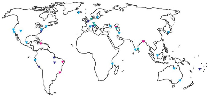
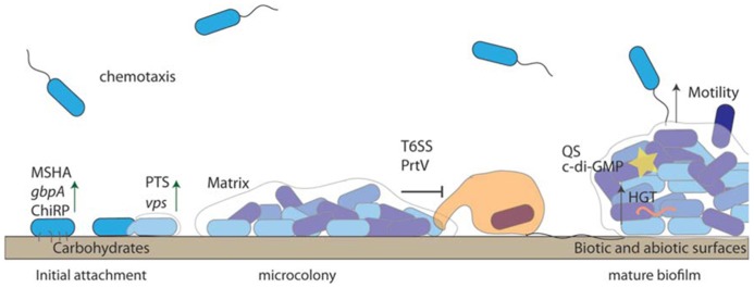
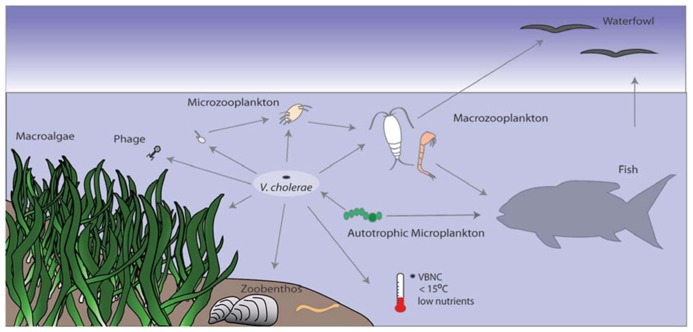
It is now well accepted that Vibrio cholerae, the causative agent of the water-borne disease cholera, is acquired from environmental sources where it persists between outbreaks of the disease. Recent advances in molecular technology have demonstrated that this bacterium can be detected in areas where it has not previously been isolated, indicating a much broader, global distribution of this bacterium outside of endemic regions. The environmental persistence of V. cholerae in the aquatic environment can be attributed to multiple intra- and interspecific strategies such as responsive gene regulation and biofilm formation on biotic and abiotic surfaces, as well as interactions with a multitude of other organisms. This review will discuss some of the mechanisms that enable the persistence of this bacterium in the environment. In particular, we will discuss how V. cholerae can survive stressors such as starvation, temperature, and salinity fluctuations as well as how the organism persists under constant predation by heterotrophic protists.

Keywords: biofilms; chitin; predation; protozoa; starvation adaptation; stress; viable but non-culturable; zooplankton.


Figures

FIGURE 1
FIGURE 1
Global distribution of Vibrio cholerae. Triangles indicate where V. cholerae was detected by molecular and/or culture-based methods. Red indicates O1/O139 detection, light blue non-O1/non-O139 detection, and dark blue did not specify. Referenced studies here are only a small fraction of the studies published for certain areas and should guide as an example. North – and Middle America: (Colwell et al., 1981; Ogg et al., 1989; Blackwell and Oliver, 2008; Lizárraga-Partida et al., 2009; Hill et al., 2011; Dickinson et al., 2013), South America: (Franco et al., 1997; Lipp et al., 2003; Leal et al., 2008; Martinelli Filho et al., 2010; Sá et al., 2012); Africa: (Taviani et al., 2008); Europe: (Andersson and Ekdahl, 2006; Covazzi Harriague et al., 2008; Kirschner et al., 2008; Vezzulli et al., 2009, Vezzulli et al., 2011; Böer et al., 2013; Cantet et al., 2013; Tall et al., 2013); Middle East: (Bakhshi et al., 2009; Grim et al., 2010; Gurbanov et al., 2011; Rashid et al., 2013); Asia Pacific: (Islam et al., 1994, 2013; Desmarchelier et al., 1995; Miyagi et al., 2003; Alam et al., 2006; Vimala et al., 2010; de Magny et al., 2011; Singh et al., 2012).
FIGURE 2
FIGURE 2
Biofilm formation enhances Vibrio cholerae persistence. V. cholerae uses chemotaxis to detect suitable surfaces for attachment. Substrate components, such as sugar concentrations of the conditioning film, play a role in the reversibly attached cells “decision” to become permanently attached. Permanent attachment is mediated by pili (ChiRP and MSHA) and outer membrane proteins such as GbpA. Biofilm formation is re-enforced through the production of VPS, which is controlled by QS (yellow star) and c-di-GMP. Anti-protozoal mechanisms such as T6SS protect surface attached bacteria. V. cholerae within biofilms undergo horizontal gene transfer (HGT), which may aid in survival.
FIGURE 3
FIGURE 3
Vibrio cholerae interactions with other organisms and the environment. V. cholerae is part of the bacterioplankton in aquatic environments. It is under predation pressure by protozoa and bacteriophage and is thus incorporated into the microbial loop. Low temperature and nutrient conditions can trigger the VBNC state, from which it resuscitates under more favorable conditions. V. cholerae can also attach to autotrophic organisms such as phytoplankton or macroalgae, which can provide a carbon source. Attachment to chitinous zooplankton and gelatinous egg masses (e.g., chironomids) provide nutrients and also facilitate HGT. Fish and birds feed on plankton or mussels that might harbor V. cholerae and can potentially spread the bacterium across long distances.

References

    1. Aagesen A, Häse C. (2012). Sequence analyses of type IV pili from Vibrio cholerae, Vibrio parahaemolyticus, and Vibrio vulnificus. Microb. Ecol. 64 509–524 10.1007/s00248-012-0021-2 - DOI - PubMed
    1. Abd H., Saeed A., Weintraub A., Nair G. B, Sandström G. (2007). Vibrio cholerae O1 strains are facultative intracellular bacteria, able to survive and multiply symbiotically inside the aquatic free-living amoeba Acanthamoeba castellanii. FEMS Microbiol. Ecol. 60 33–39 10.1111/j.1574-6941.2006.00254.x - DOI - PubMed
    1. Abd H., Saeed A., Weintraub A, Sandström G. (2009). Vibrio cholerae O139 requires neither capsule nor LPS O side chain to grow inside Acanthamoeba castellanii. J. Med. Microbiol. 58 125–131 10.1099/jmm.0.004721-0 - DOI - PMC - PubMed
    1. Abd H., Shanan S., Saeed A, Sandström G. (2011). Survival of Vibrio cholerae inside Acanthamoeba and detection of both microorganisms from natural water samples may point out the amoeba as a protozoal host for V. cholerae. J. Bacteriol. Parasitol. 4 109
    1. Abd H., Weintraub A, Sandström G. (2004). Interaction between Vibrio cholerae and Acanthamoeba castellanii. Microb. Ecol. Health Dis. 16 51–57 10.1080/08910600410029190 - DOI

LinkOut - more resources