

SEM evaluation of pulp reaction to different pulp capping materials in dog's teeth
- PMID: 24379876
- PMCID: PMC3874105
SEM evaluation of pulp reaction to different pulp capping materials in dog's teeth
Abstract
Introduction: This investigation evaluates the effects of mineral trioxide aggregate (MTA), calcium hydroxide (CH) and calcium enriched mixture (CEM) as pulp capping materials on dental pulp tissues.
Materials and methods: The experimental procedures were performed on eighteen intact dog canine teeth. The pulps were exposed. Cavities were randomly filled with CEM, MTA, or CH followed by glass ionomer filling. After 2 months, animals were sacrificed, each tooth was sectioned into halves, and the interface between each capping material and pulp tissue was evaluated by scanning electron microscope (SEM) in profile view of the specimens.
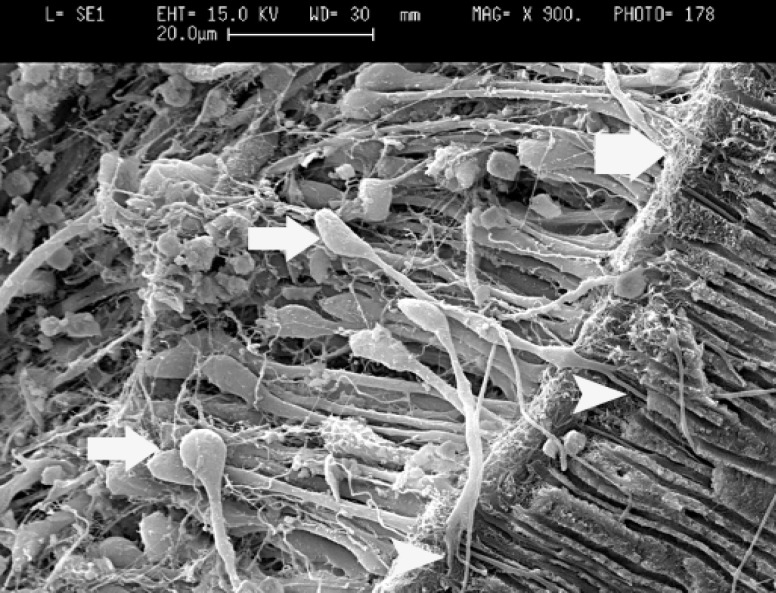
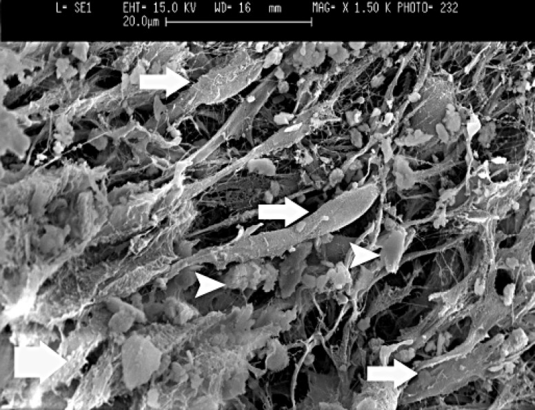
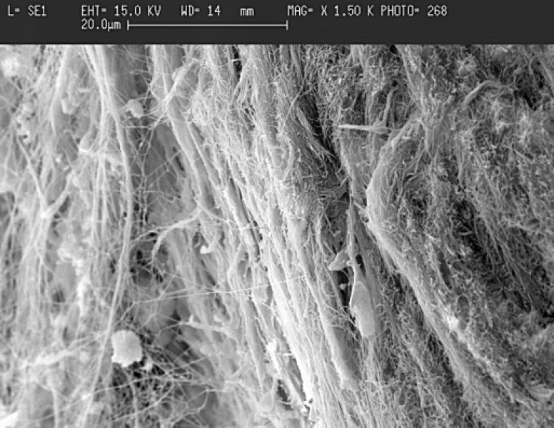
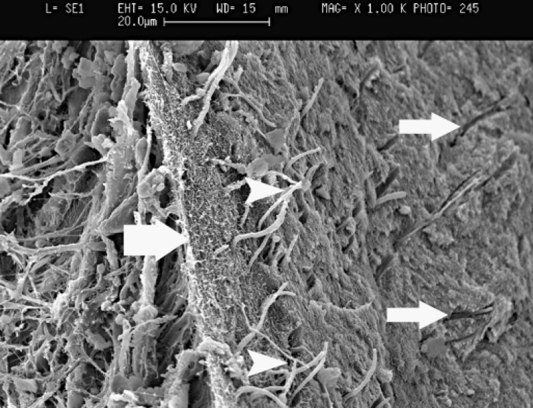
Results: Dentinal bridge formation as the most characteristic reaction was resulted from SEM observation in all examined groups. Odontoblast-like cells were formed and create dens collagen network, which was calcified gradually by deposition of calcosphirit structures to form newly dentinal bridge.
Conclusion: Based on the results of this in vivo study, it was concluded that these test materials are able to produce calcified tissue in underlying pulp in the case of being used as a pulp capping agent. Additionally, it appears that CEM has the potential to be used as a direct pulp capping material during vital pulp therapy.
Keywords: Calcium enriched mixture; Calcium hydroxide; MTA; Pulp capping agent; SEM.
Figures
References
-
- Bakland LK. Endodontic considerations in dental trauma. In: Ingle JI, Bakland LK, editors. Endodontics. 5th ed. Toronto: BC Decker; 2002. pp. 795–844.
-
- Mjör IA, Dahl E, Cox CF. Healing of pulp exposures: an ultrastructural study. J Oral Pathol Med. 1991;20:496–501. - PubMed
-
- Murray PE, Hafez AA, Smith AJ, Cox CF. Hierarchy of pulp capping and repair activities responsible for dentin bridge formation. Am J Dent. 2002;15:236–43. - PubMed
-
- Hess W. Pulp amputation as a method of treating root canals. D Items Int. 1929;51:596.
-
- Dummett CO, Kopel HM. Pediatric endodo-ntics. In: Ingle JI, Bakland LK, editors. Endodontics. 5th ed. Toronto: BC Decker; 2002. pp. 861–902.
LinkOut - more resources
Full Text Sources