

Classification of diabetic retinopathy and diabetic macular edema
- PMID: 24379919
- PMCID: PMC3874488
- DOI: 10.4239/wjd.v4.i6.290
Classification of diabetic retinopathy and diabetic macular edema
Abstract
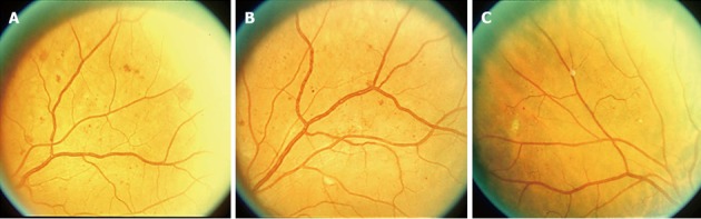
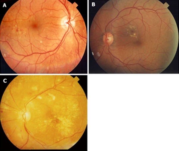
The global incidence and prevalence of diabetes mellitus (DM) have reached epidemic proportions. Estimates indicate that more than 360 million people will be affected by DM by 2030. All of these individuals will be at risk of developing diabetic retinopathy (DR). It is extremely important to categorize, classify and stage the severity of DR in order to establish adequate therapy. With proper management more than 90% of cases of visual loss can be prevented. The purpose of the current paper is to review the classification of DR with a special emphasis on the International Clinical Disease Severity Scale for DR. This new classification is simple to use, easy to remember and based on scientific evidence. It does not require specialized examinations such as optical coherence tomography or fluorescein angiography. It is based on clinical examination and applying the Early Treatment of Diabetic Retinopathy Study 4:2:1 rule.
Keywords: Diabetic macular edema; Diabetic macular edema classification; Diabetic retinopathy; Diabetic retinopathy classification; International clinical disease severity scale.
Figures
References
-
- Wild S, Roglic G, Green A, Sicree R, King H. Global prevalence of diabetes: estimates for the year 2000 and projections for 2030. Diabetes Care. 2004;27:1047–1053. - PubMed
-
- Engerman RL, Kern TS. Retinopathy in animal models of diabetes. Diabetes Metab Rev. 1995;11:109–120. - PubMed
-
- Klein R, Klein BE, Moss SE, Cruickshanks KJ. The Wisconsin Epidemiologic Study of diabetic retinopathy. XIV. Ten-year incidence and progression of diabetic retinopathy. Arch Ophthalmol. 1994;112:1217–1228. - PubMed
-
- Klein R, Klein BE, Moss SE. Visual impairment in diabetes. Ophthalmology. 1984;91:1–9. - PubMed
-
- Williams R, Airey M, Baxter H, Forrester J, Kennedy-Martin T, Girach A. Epidemiology of diabetic retinopathy and macular oedema: a systematic review. Eye (Lond) 2004;18:963–983. - PubMed
Publication types
LinkOut - more resources
Full Text Sources
Other Literature Sources
Medical
