

(68)Ga-labeled superparamagnetic iron oxide nanoparticles (SPIONs) for multi-modality PET/MR/Cherenkov luminescence imaging of sentinel lymph nodes
- PMID: 24380046
- PMCID: PMC3867730
(68)Ga-labeled superparamagnetic iron oxide nanoparticles (SPIONs) for multi-modality PET/MR/Cherenkov luminescence imaging of sentinel lymph nodes
Abstract
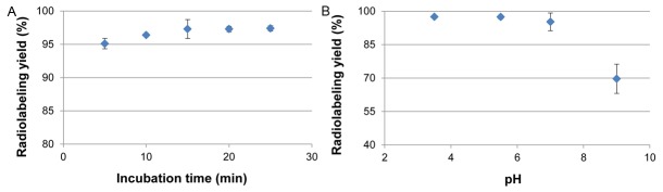
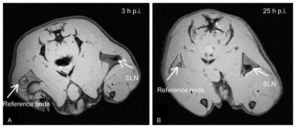
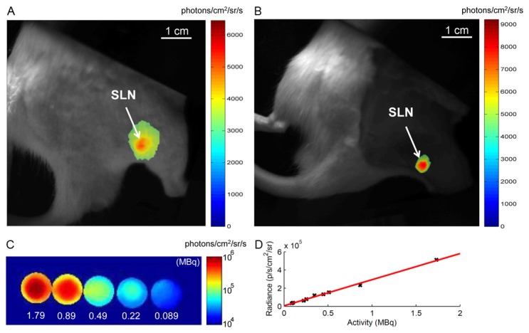
The aim of this study was to develop (68)Ga-SPIONs for use as a single contrast agent for dynamic, quantitative and high resolution PET/MR imaging of Sentinel Lymph Node (SLN). In addition (68)Ga enables Cherenkov light emission which can be used for optical guidance during resection of SLN. SPIONs were labeled with (68)Ga in ammonium acetate buffer, pH 5.5. The labeling yield and stability in human serum were determined using instant thin layer chromatography. An amount of 0.07-0.1 mL (~5-10 MBq, 0.13 mg Fe) of (68)Ga-SPIONs was subcutaneously injected in the hind paw of rats. The animals were imaged at 0-3 h and 25 h post injection with PET/CT, 9.4 T MR and CCDbased Cherenkov optical systems. A biodistribution study was performed by dissecting and measuring the radioactivity in lymph nodes, kidneys, spleen, liver and the injection site. The labeling yield was 97.3 ± 0.05% after 15 min and the (68)Ga-SPIONs were stable in human serum. PET, MR and Cherenkov luminescence imaging clearly visualized the SLN. Biodistribution confirmed a high uptake of the (68)Ga-SPIONs within the SLN. We conclude that generator produced (68)Ga can be labeled to SPIONs. Subcutaneously injected (68)Ga-SPIONs can enhance the identification of the SLNs by combining sensitive PET and high resolution MR imaging. Clinically, hybrid PET/MR cameras are already in use and (68)Ga-SPIONs have a great potential as a single-dose, tri-modality agent for diagnostic imaging and potential Cherenkov luminescent guided resection of SLN.
Keywords: 68Ga; Cherenkov imaging; PET/MR imaging; lymphatics; sentinel lymph node (SLN); superparamagnetic iron oxide nanoparticle (SPION).
Figures


Similar articles
-
99mTc-labeled superparamagnetic iron oxide nanoparticles for multimodality SPECT/MRI of sentinel lymph nodes.J Nucl Med. 2012 Mar;53(3):459-63. doi: 10.2967/jnumed.111.092437. Epub 2012 Feb 9. J Nucl Med. 2012. PMID: 22323777
-
Heterogeneous Distribution and Absorbed Dose of Radiolabeled Nanoparticles and Colloids in Sentinel Lymph Nodes.Cancer Biother Radiopharm. 2025 May;40(4):254-262. doi: 10.1089/cbr.2024.0111. Epub 2025 Feb 26. Cancer Biother Radiopharm. 2025. PMID: 40000017
-
Development of a Hybrid Nanoprobe for Triple-Modality MR/SPECT/Optical Fluorescence Imaging.Diagnostics (Basel). 2014 Mar 10;4(1):13-26. doi: 10.3390/diagnostics4010013. Diagnostics (Basel). 2014. PMID: 26852675 Free PMC article.
-
[Magnetic Sentinel Lymph Node Detection in Prostate Cancer after intraprostatic Injection of Superparamagnetic Iron Oxide Nanoparticles].Aktuelle Urol. 2017 Apr;48(2):132-139. doi: 10.1055/s-0042-121338. Epub 2017 Mar 21. Aktuelle Urol. 2017. PMID: 28324908 Review. German.
-
New Frontiers in Molecular Imaging with Superparamagnetic Iron Oxide Nanoparticles (SPIONs): Efficacy, Toxicity, and Future Applications.Nucl Med Mol Imaging. 2020 Apr;54(2):65-80. doi: 10.1007/s13139-020-00635-w. Epub 2020 Feb 8. Nucl Med Mol Imaging. 2020. PMID: 32377258 Free PMC article. Review.
Cited by
-
Amplification of Cerenkov luminescence using semiconducting polymers for cancer theranostics.Adv Funct Mater. 2023 Aug 15;33(33):2302777. doi: 10.1002/adfm.202302777. Epub 2023 May 1. Adv Funct Mater. 2023. PMID: 37942189 Free PMC article.
-
Cerenkov luminescence imaging (CLI) for image-guided cancer surgery.Clin Transl Imaging. 2016;4(5):353-366. doi: 10.1007/s40336-016-0183-x. Epub 2016 May 24. Clin Transl Imaging. 2016. PMID: 27738626 Free PMC article. Review.
-
Preparation and PET/CT imaging of implant directed 68Ga-labeled magnetic nanoporous silica nanoparticles.J Nanobiotechnology. 2023 Aug 17;21(1):270. doi: 10.1186/s12951-023-02041-8. J Nanobiotechnology. 2023. PMID: 37592318 Free PMC article.
-
Radionuclide generators: the prospect of availing PET radiotracers to meet current clinical needs and future research demands.Am J Nucl Med Mol Imaging. 2019 Feb 15;9(1):30-66. eCollection 2019. Am J Nucl Med Mol Imaging. 2019. PMID: 30911436 Free PMC article. Review.
-
Multimodality Imaging Agents with PET as the Fundamental Pillar.Angew Chem Int Ed Engl. 2019 Feb 25;58(9):2570-2579. doi: 10.1002/anie.201806853. Epub 2018 Dec 11. Angew Chem Int Ed Engl. 2019. PMID: 29968300 Free PMC article. Review.
References
-
- Alitalo K, Tammela T, Petrova TV. Lymphangiogenesis in development and human disease. Nature. 2005;438:946–953. - PubMed
-
- Civantos FJ, Zitsch RP, Schuller DE, Agrawal A, Smith RB, Nason R, Petruzelli G, Gourin CG, Wong RJ, Ferris RL, El Naggar A, Ridge JA, Paniello RC, Owzar K, McCall L, Chepeha DB, Yarbrough WG, Myers JN. Sentinel lymph node biopsy accurately stages the regional lymph nodes for T1-T2 oral squamous cell carcinomas: results of a prospective multi-institutional trial. J. Clin. Oncol. 2010;28:1395–1400. - PMC - PubMed
-
- Wong SL, Balch CM, Hurley P, Agarwala SS, Akhurst TJ, Cochran A, Cormier JN, Gorman M, Kim TY, McMasters KM, Noyes RD, Schuchter LM, Valsecchi ME, Weaver DL, Lyman GH. Sentinel lymph node biopsy for melanoma: American Society of Clinical Oncology and Society of Surgical Oncology joint clinical practice guideline. J. Clin. Oncol. 2012;30:2912–2918. - PMC - PubMed
-
- McMasters KM, Tuttle TM, Carlson C, Brown CM, Noyes RD, Glaser RL, Vennekotter DJ, Turk PS, Tate PS, Sardi A, Cerrito PB, Edwards MJ. Sentinel lymph node biopsy for breast cancer a suitable alternative to routine axillary dissection in multi-institutional practice when optimal technique is used. J. Clin. Oncol. 2000;18:2560–2566. - PubMed
-
- Balch CM, Gershenwald JE, Soong SJ, Thompson JF, Atkins MB, Byrd DR, Buzaid AC, Cochran AJ, Coit DG, Ding S, Eggermont AM, Flaherty KT, Gimotty PA, Kirkwood JM, McMasters KM, Mihm MC Jr, Morton DL, Ross MI, Sober AJ, Sondak VK. Final version of 2009 AJCC melanoma staging and classification. J. Clin. Oncol. 2009;27:6199–6206. - PMC - PubMed
LinkOut - more resources
Full Text Sources
Other Literature Sources