Review
. 2013 Dec 31;66(1):334-95.
doi: 10.1124/pr.112.007336. Print 2014.

Computational methods in drug discovery

Affiliations
Review

Computational methods in drug discovery

Gregory Sliwoski et al. Pharmacol Rev. .

Abstract

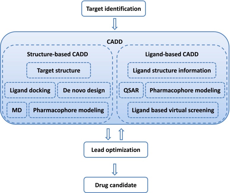
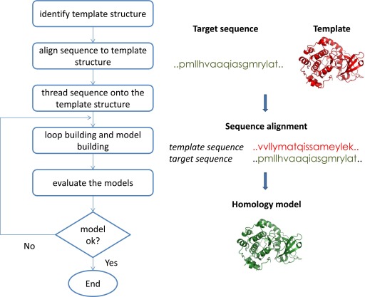
Computer-aided drug discovery/design methods have played a major role in the development of therapeutically important small molecules for over three decades. These methods are broadly classified as either structure-based or ligand-based methods. Structure-based methods are in principle analogous to high-throughput screening in that both target and ligand structure information is imperative. Structure-based approaches include ligand docking, pharmacophore, and ligand design methods. The article discusses theory behind the most important methods and recent successful applications. Ligand-based methods use only ligand information for predicting activity depending on its similarity/dissimilarity to previously known active ligands. We review widely used ligand-based methods such as ligand-based pharmacophores, molecular descriptors, and quantitative structure-activity relationships. In addition, important tools such as target/ligand data bases, homology modeling, ligand fingerprint methods, etc., necessary for successful implementation of various computer-aided drug discovery/design methods in a drug discovery campaign are discussed. Finally, computational methods for toxicity prediction and optimization for favorable physiologic properties are discussed with successful examples from literature.


Figures

Fig. 1.
Fig. 1.
Identical lead compounds are discovered in a traditional high-throughput screen and structure-based virtual high-throughput screen. I, X-ray crystal structures of 1 and 18 bound to the ATP-binding site of the TβR-I kinase domain discovered using traditional high-throughput screening. Compound 1, shown as the thinner wire-frame is the original hit from the HTS and is identical to that which was discovered using virtual screening. Compound 18 is a higher affinity compound after lead optimization. II, X-ray crystal structure of compound HTS466284 bound to the TβRI active site. This compound is identical to compound 1 in I but was discovered using structure-based virtual high-throughput screening.
Fig. 2.
Fig. 2.
CADD in drug discovery/design pipeline. A therapeutic target is identified against which a drug has to be developed. Depending on the availability of structure information, a structure-based approach or a ligand-based approach is used. A successful CADD campaign will allow identification of multiple lead compounds. Lead identification is often followed by several cycles of lead optimization and subsequent lead identification using CADD. Lead compounds are tested in vivo to identify drug candidates .
Fig. 3.
Fig. 3.
Steps in homology model building process.
Fig. 4.
Fig. 4.
Discovery of novel binding trench in HIV-1 IN. Ligand in green is similar to the crystal structure binding pose while the one in yellow is in the novel trench. Adapted from Schames et al. (2004).
Fig. 5.
Fig. 5.
Lead compound obtained through virtual screening of a library of compounds against M1 mAChR. Adapted from Budzik et al. (2010).
Fig. 6.
Fig. 6.
Potent inhibitors of protein kinase CK2—ellagic acid and quinalizarin.
Fig. 7.
Fig. 7.
Outline of discovery process of novel family of PPAR-γ partial agonists. A, Conformation of compound 6 bound to active site of PPAR-γ was used as a pharmacophore. B, The bound conformation of compound 6 was used to screen the compound library. Compound 7 identified as a hit in the compound library screen. The binding mode of compound 7 obtained through docking study was used to define a core structure that was used for further similarity search which identified compound 1 as a potent agonist of PPAR-γ. From Lu et al. (2006).
Fig. 8.
Fig. 8.
Optimization of compound 8 for selective binding to 5-HT1A over α1-adrenergic receptor. 1a and 1b, Interactions of compound 8 with 5-HT and α1, respectively. The dotted box represents the structural differences between the two target molecules. The authors leveraged this difference between the protein molecules to design a virtual analog of compound 8, identified as 20h. 2b and 2b, docking models of 20h into 5-HT and α1. These docking modes indicate that the piperazine atom and aspartic acid interaction is maintained for 20h-5-HT complex and not for 20h-α1 complex. An optimization strategy based on this observation was used to design the novel agonist PRX-00023 for treatment of anxiety and depression. Adapted from Becker et al. (2006).
Fig. 9.
Fig. 9.
A comparison of the hERG binding modes of compounds 8 and 20d. Shown are a detailed 3D view of the binding of compounds 8 in the hERG pore, as well as two schematic views of the binding of compounds 8 and 20d next to each other. The four main interaction regions are highlighted in all views: an aromatic region formed by the four Tyr652 residues, a K+ pocket, an aromatic region formed by the four Phe656 residues, and a polar region formed by four Ser660 residues (shown only schematically).
Fig. 10.
Fig. 10.
Carboxynoxolone and 10j2. Overlap of carenoxolone (yellow) and urea 10j2 (green) in binding site of 11β-HSD1.
Fig. 11.
Fig. 11.
Evolution of the design of novel HCV helicase inhibitor.
Fig. 12.
Fig. 12.
Extracting common pharmacophores of LTA4H-h and human-PLA2. Cyan spheres depict hydrophobic centers, red spheres represent H-bond acceptor while yellow spheres stand for feature that coordinates with a metal. Adapted from Wei et al. (2008).
Fig. 13.
Fig. 13.
(A) A reported inhibitor of LTA4H-h. (B) Compound 11.
Fig. 14.
Fig. 14.
Design strategy for inhibitors of p38 MAPK. (A) Key interactions of BIRB-796 inhibitor with MAPK. (B) A fragment linking strategy to link two seed structures was applied using LigBuilder. A tert-butyl phenyl fragment was used in the first pocket, whereas a carbonyl fragment was used to access the hydrogen bond with Met109 in the second site. An N-formyl group was attached to the first seed fragment to access hydrogen bonds with Glu71 and Asp168. (C) General structure of optimized structures which showed potent activity. (D) R group for compound 28, which showed IC50 value of 83 nM. Adapted from Cogan et al. (2008).
Fig. 15.
Fig. 15.
(A) Chemical structure of SKLB1002. (B) SKLB1002 is docked into the active site of VEGFR2, showing interactions between SKLB1002 and VEGFR2 by using the in silico model. (C) A 2D interaction map of SKLB1002 and VEGFR2. Adapted from Zhang et al. (2011).
Fig. 16.
Fig. 16.
QSAR-based virtual screening of mGlu5 negative allosteric modulators yields lead compounds that contain substructure combinations taken across several known actives used for model generation. Adapted from Mueller et al. (2012).
Fig. 17.
Fig. 17.
SR13668, an anticancer therapeutic was discovered using ligand-based pharmacophore screening based on active components of indole-3-carbinol. Adapted from Chao et al. (2007).
Fig. 18.
Fig. 18.
(I, A) Novel HIV-1 Integrase inhibitor using ligand-based virtual screening with a pharmacophore model of quinolone 3-carboxylic acid IN inhibitors [from Dayam et al. (2008)]. (B) Pharmacophore query generated from the quinolone 3-carboxylic acid IN inhibitors accompanied with an overlay onto a known HIV-1 integrase inhibitor. Features are color-coded, and their 3D arrangement/distances are shown in angstroms. Green sphere represent H-bond acceptor regions, blue spheres represent negatively ionizable regions, and cyan spheres represent hydrophobic aromatic regions. (II) Pharmacophore query overlayed with 3 potent hits from the ligand-based virtual screen: compounds 8 (A), 9 (B), and 17 (C).
Fig. 19.
Fig. 19.
Ripphausen et al. (2010) report that ligand-based computationally approaches yield compounds with higher affinity than structure-based computationally approaches. Adapted from Ripphausen et al. (2010).

References

    1. Abagyan R, Lee WH, Raush E, Budagyan L, Totrov M, Sundstrom M, Marsden BD. (2006) Disseminating structural genomics data to the public: from a data dump to an animated story. Trends Biochem Sci 31:76–78 - PubMed
    1. Abagyan R, Totrov M,, Kuznetsov D. (1994) ICM - a new method for protein modeling and design–applications to docking and structure prediction from the distorted native conformation. J Comput Chem 15:488–506
    1. Abrams CF, Vanden-Eijnden E. (2010) Large-scale conformational sampling of proteins using temperature-accelerated molecular dynamics. Proc Natl Acad Sci USA 107:4961–4966 - PMC - PubMed
    1. Accelrys (2013) Accelrys metabolite. Available from http://accelrys.com/products/databases/bioactivity/metabolite.html
    1. Acharya C, Coop A, Polli JE, Mackerell AD., Jr (2011) Recent advances in ligand-based drug design: relevance and utility of the conformationally sampled pharmacophore approach. Curr Comput Aided Drug Des 7:10–22 - PMC - PubMed

Publication types