

The Repertoires of Peptides Presented by MHC-II in the Thymus and in Peripheral Tissue: A Clue for Autoimmunity?
- PMID: 24381570
- PMCID: PMC3865459
- DOI: 10.3389/fimmu.2013.00442
The Repertoires of Peptides Presented by MHC-II in the Thymus and in Peripheral Tissue: A Clue for Autoimmunity?
Abstract
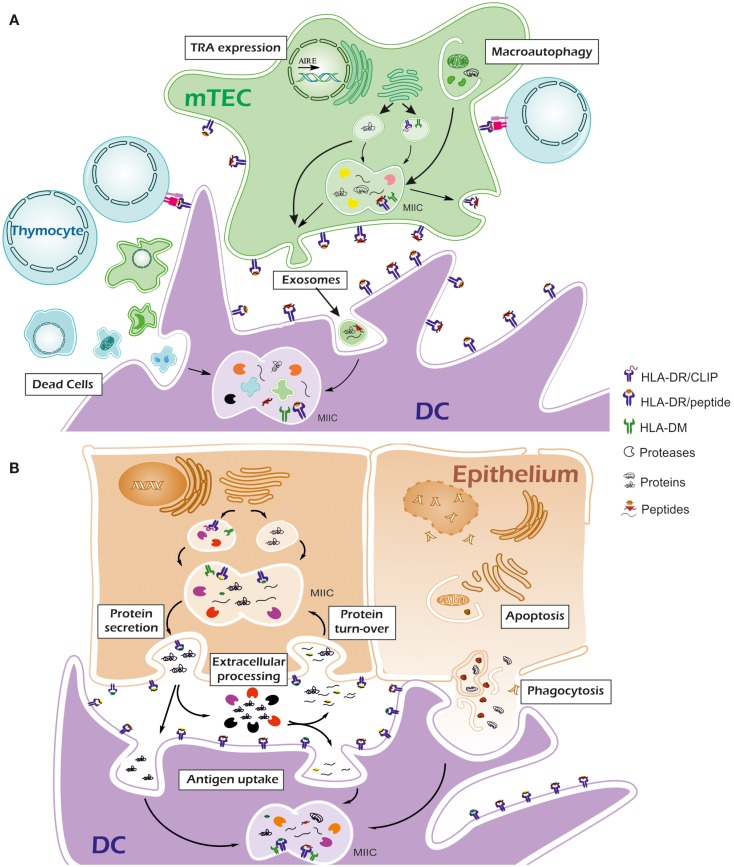
T-cell tolerance to self-antigens is established in the thymus through the recognition by developing thymocytes of self-peptide-MHC complexes and induced and maintained in the periphery. Efficient negative selection of auto-reactive T cells in the thymus is dependent on the in situ expression of both ubiquitous and tissue-restricted self-antigens and on the presentation of derived peptides. Weak or inadequate intrathymic expression of self-antigens increases the risk to generate an autoimmune-prone T-cell repertoire. Indeed, even small changes of self-antigen expression in the thymus affect negative selection and increase the predisposition to autoimmunity. Together with other mechanisms, tolerance is maintained in the peripheral lymphoid organs via the recognition by mature T cells of a similar set of self-peptides in homeostatic conditions. However, non-lymphoid peripheral tissue, where organ-specific autoimmunity takes place, often have differential functional processes that may lead to the generation of epitopes that are absent or non-presented in the thymus. These putative differences between peptides presented by MHC molecules in the thymus and in peripheral tissues might be a major key to the initiation and maintenance of autoimmune conditions.
Keywords: MHC-II; human; peptide repertoire; peripheral tissues; thymus.
Figures
Similar articles
-
MHC and T cell development.Rev Immunogenet. 1999;1(1):91-104. Rev Immunogenet. 1999. PMID: 11256575 Review.
-
A novel pathway of presentation by class II-MHC molecules involving peptides or denatured proteins important in autoimmunity.Mol Immunol. 2013 Sep;55(2):166-8. doi: 10.1016/j.molimm.2012.10.024. Epub 2012 Nov 27. Mol Immunol. 2013. PMID: 23200226 Free PMC article. Review.
-
Thymic commitment of regulatory T cells is a pathway of TCR-dependent selection that isolates repertoires undergoing positive or negative selection.Curr Top Microbiol Immunol. 2005;293:43-71. doi: 10.1007/3-540-27702-1_3. Curr Top Microbiol Immunol. 2005. PMID: 15981475 Review.
-
Thymic T-cell tolerance of neuroendocrine functions: physiology and pathophysiology.Cell Mol Biol (Noisy-le-grand). 2001 Feb;47(1):179-88. Cell Mol Biol (Noisy-le-grand). 2001. PMID: 11292253 Review.
-
Self major histocompatibility complex class I antigens expressed solely in lymphoid cells do not induce tolerance in the CD4+ T cell compartment.J Exp Med. 1996 Oct 1;184(4):1573-8. doi: 10.1084/jem.184.4.1573. J Exp Med. 1996. PMID: 8879232 Free PMC article.
Cited by
-
Autoimmune BSEP disease: disease recurrence after liver transplantation for progressive familial intrahepatic cholestasis.Clin Rev Allergy Immunol. 2015 Jun;48(2-3):273-84. doi: 10.1007/s12016-014-8457-4. Clin Rev Allergy Immunol. 2015. PMID: 25342496 Review.
-
Thinking bedside at the bench: the NOD mouse model of T1DM.Nat Rev Endocrinol. 2015 May;11(5):308-14. doi: 10.1038/nrendo.2014.236. Epub 2015 Jan 27. Nat Rev Endocrinol. 2015. PMID: 25623120 Free PMC article. Review.
-
Potential roles of neutrophils in maintaining the health and productivity of dairy cows during various physiological and physiopathological conditions: a review.Immunol Res. 2019 Feb;67(1):21-38. doi: 10.1007/s12026-019-9064-5. Immunol Res. 2019. PMID: 30644032 Review.
-
The Protective Role of HLA-DRB1(∗)13 in Autoimmune Diseases.J Immunol Res. 2015;2015:948723. doi: 10.1155/2015/948723. Epub 2015 Oct 29. J Immunol Res. 2015. PMID: 26605347 Free PMC article.
-
Human thymic epithelial primary cells produce exosomes carrying tissue-restricted antigens.Immunol Cell Biol. 2015 Sep;93(8):727-34. doi: 10.1038/icb.2015.33. Epub 2015 Mar 17. Immunol Cell Biol. 2015. PMID: 25776846 Free PMC article.
References
Publication types
LinkOut - more resources
Full Text Sources
Other Literature Sources
Research Materials