. 2013 Jul;3(4):384-401.
doi: 10.1002/brb3.135. Epub 2013 Apr 24.

An fMRI investigation of delay discounting in patients with schizophrenia

Affiliations

An fMRI investigation of delay discounting in patients with schizophrenia

Kathy Burton Avsar et al. Brain Behav. 2013 Jul.

Abstract

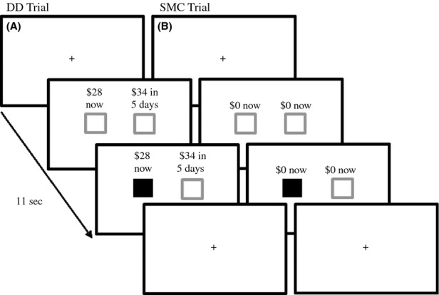
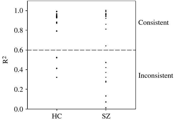
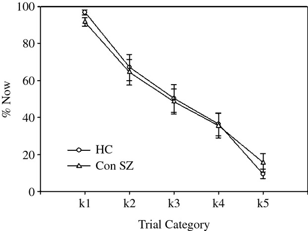
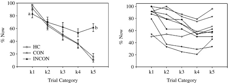
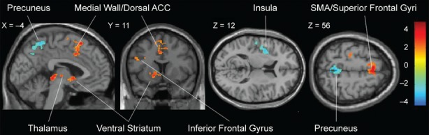
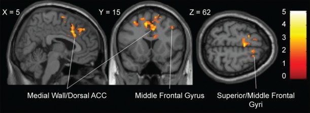
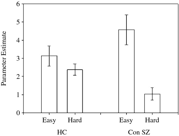
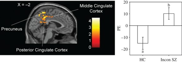
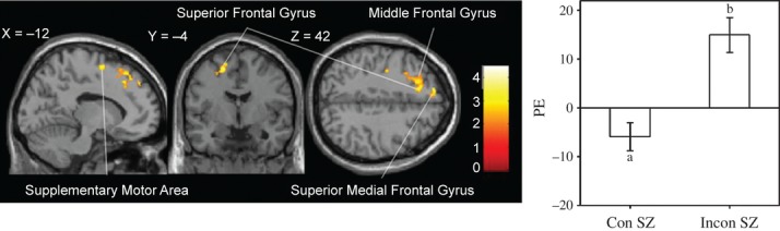
Schizophrenia (SZ) is associated with a reduced ability to set meaningful goals to reach desired outcomes. The delay-discounting (DD) task, in which one chooses between sooner smaller and later larger rewards, has proven useful in revealing executive function and reward deficits in various clinical groups. We used fMRI in patients with SZ and healthy controls (HC) to compare brain activation during performance of a DD task. Prior to the neuroimaging session, we obtained each participant's rate of DD, k, on a DD task and used it to select a version of the DD task for each participant's fMRI session. Because of the importance of comparing fMRI results from groups matched on performance, we used a criterion value of R (2) > 0.60 for response consistency on the DD task to analyze fMRI activation to DD task versus control trials from consistent SZ (n = 14) and consistent HC (n = 14). We also compared activation between the groups on contrasts related to trial difficulty. Finally, we contrasted the inconsistent SZ (n = 9) with the consistent HC and consistent SZ; these results should be interpreted with caution because of inconsistent SZ's aberrant performance on the task. Compared with consistent HC, consistent SZ showed reduced activation to DD task versus control trials in executive function and reward areas. In contrast, consistent SZ showed more activation in the precuneus and posterior cingulate, regions of the default mode network (DMN) that are typically deactivated during tasks, and in the insula, a region linked to emotional processing. Furthermore, consistent SZ had abnormal activation of lateral and medial frontal regions in relation to trial difficulty. These results point to disruption of several neural networks during decision making, including the executive, reward, default mode, and emotional networks, and suggest processes that are impaired during decision making in schizophrenia.

Keywords: Delay discounting; executive function; intertemporal; reward; schizophrenia; subjective value.


Figures

Figure 1
Figure 1
Delay-discounting (DD) task. (A) DD task trial; (B) sensorimotor control (SMC) trial. All trials were 11 sec in duration, with the initial fixation cross presented for 2, 4, or 6 sec, followed by two gray boxes paired with (A) the choice of an immediate or a delayed hypothetical monetary reward ($28 now or $34 in 5 days; a trial k of 0.041) or (B) the no-choice option. Participants had the remainder of the 11 sec trial (9, 7, or 5 sec) to indicate their preference by pressing a button on the side corresponding to their choice. The box under the choice turned green, indicating the response selection. A return of the fixation cross indicated the start of the next trial.
Figure 2
Figure 2
Individual model fit (R2) values during estimation of k values for healthy controls (HC) and patients with schizophrenia (SZ). The line at 0.60 indicates the minimum R2 value that was used to define consistent performance.
Figure 3
Figure 3
Mean (± standard error) for percentage of Now (%Now) choices as a function of the five trial k's for the consistent HC and consistent patients with schizophrenia (Con SZ).
Figure 4
Figure 4
Mean (± standard error) of response times across the five trial categories during the scanning session for the consistent healthy controls (HC) and consistent patients (Con SZ). *P < 0.05 between groups.
Figure 5
Figure 5
Mean (± standard error) percentage of Now (%Now) choices as a function of the five trial k's for the consistent healthy controls (HC), consistent SZ, and inconsistent SZ (INCON) (left) and for individual inconsistent SZ (n = 9; right). a, P = 0.005 for inconsistent SZ versus HC and 0.07 versus consistent SZ; b, P < 0.001 for inconsistent SZ versus HC and versus consistent SZ.
Figure 6
Figure 6
Between-group results for activation to task>SMC trials revealed more activation in controls (red) in frontoparietal areas, including inferior frontal gyrus and medial areas of the prefrontal cortex, and subcortically in the striatum and thalamus; x, y, and z are MNI coordinates. Patients (blue) had more activation than controls in the precuneus and insula. P < 0.05, false discovery rate (FDR) corrected for cluster extent. Bar at right represents t values.
Figure 7
Figure 7
Between-group results for activation to hard>easy trials revealed an interaction between difficulty and group. For controls>consistent patients, the contrast is hard>easy; for consistent patients>controls, the contrast is easy>hard. P < 0.05, FDR-corrected. Conventions as in Figure 6.
Figure 8
Figure 8
Mean (± standard error) parameters estimates extracted from the each participant's contrast maps for hard trials and easy trials using a functionally defined composite mask for the between-group results for hard versus easy trials. HC, healthy controls; Con SZ, consistent SZ.
Figure 9
Figure 9
Between-group fMRI results for activation in inconsistent patients (n = 9) when compared with consistent controls (n = 14) to task>SMC trials for the largest, medial cluster activated. Left, the sagittal brain section shows greater activation occurred in inconsistent SZ when compared with consistent controls on the medial wall (medial wall/parietal/occipital cluster size 3048 of Table 4); in particular, in the precuneus and posterior and middle cingulate cortex. Voxel-level intensity threshold uncorrected P < 0.05, with a minimum cluster size to maintain a FDR = 0.05. No regions were more activated in the consistent controls than in the inconsistent SZ. Right, mean (± standard error) parameter estimates (PE) extracted for the functionally defined mask of group differences in inconsistent SZ (task>SMC) > consistent controls (task>SMC) for the same medial wall cluster. a, P = 0.043 versus 0; b, P = 0.076 versus 0.
Figure 10
Figure 10
Left, the brain section shows between-group fMRI results for activation to task>SMC trials. More activation occurred in inconsistent SZ (n = 9) when compared with consistent SZ (n = 14) in the supplementary motor area, superior frontal, and superior medial frontal gyri. Right, mean (± standard error) parameter estimates (PE) extracted for the functionally defined mask of group difference for inconsistent patients (task>SMC) > consistent patients (task>SMC) for the cluster of Table 4 with the peak voxel at MNI coordinates −33, 24, and 46 in the middle frontal gyrus. a, P = 0.059 versus 0; b, P = 0.003 versus 0. Other conventions as in Figure 9.

References

    1. Ahn WY, Rass O, Fridberg DJ, Bishara AJ, Forsyth JK, Breier A, et al. Temporal discounting of rewards in patients with bipolar disorder and schizophrenia. J. Abnorm. Psychol. 2011;120:911–921. - PMC - PubMed
    1. Ainslie G. Specious reward: a behavioral theory of impulsiveness and impulse control. Psychol. Bull. 1975;82:463–496. - PubMed
    1. American Psychiatric Association. American Psychiatric Association. Task Force on DSM-IV. Diagnostic and statistical manual of mental disorders: DSM-IV-TR. 4th ed. Washington, DC: American Psychiatric Association; 2000.
    1. Ashburner J. A fast diffeomorphic image registration algorithm. NeuroImage. 2007;38:95–113. - PubMed
    1. Avsar KB, Stoeckel LE, Bolding MS, White DM, Tagamets MA, Holcomb HH, et al. Aberrant visual circuitry associated with normal spatial match-to-sample accuracy in schizophrenia. Psychiatry Res. 2011;193:138–143. - PubMed

LinkOut - more resources