

Fungus cerebri (brain fungus): a rare complication of mastoidectomy
- PMID: 24381920
- PMCID: PMC3585555
- DOI: 10.1007/s12070-012-0601-y
Fungus cerebri (brain fungus): a rare complication of mastoidectomy
Abstract
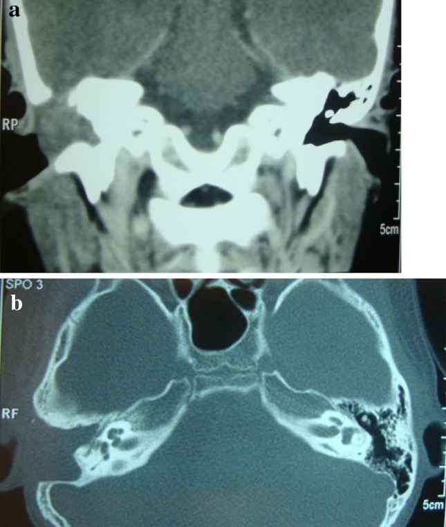
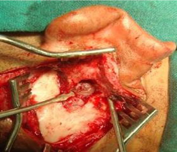
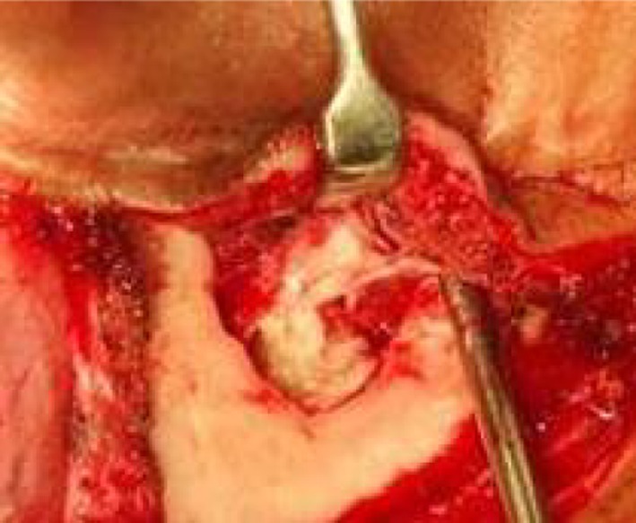
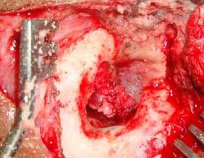
Fungus cerebri is a relatively rare disease. The various reasons attributed to such pathology are, long standing mastoiditis, previous temporal lobe fracture, spontaneous herniation and most important common cause is post operative to mastoidectomy. The diagnosis is mainly clinical and supplemented by imaging studies. The commonly herniated part is the temporal lobe, but cerebellar herniation are also reported Different surgical modalities are used in managing this condition. Surgical approaches in the treatment of brain herniation into the mastoid or middle ear are, neurosurgical, otosurgical and combined. A case of fungus cerebri complicating mastoidectomy is presented and the pathogenesis is discussed.
Keywords: Brain fungus; Fungus cerebri; Masoidectomy.
Figures


References
-
- Gluckman J. Fungus cerebri: an unusual complication of mastoidectomy. S Afr Med J. 1975;49(46):1933–1934. - PubMed
-
- Jackson CG, Pappas DG Jr, Manolidis S, Glasscock ME, Von Doersten PG, Carl R Hampf CR, et al. Brain herniation into the middle ear and mastoid: concepts in diagnosis and surgical management. Am J Otolaryngol. 1997;18:198–206. - PubMed
-
- Ramalingam KK, RaviRamalingam, Sreenivasa Murthy TM, Chandrakala GR, UttamAgarwal An unusual case of brain fungus. Orissa J Otolaryngol Head & Neck Surg. 2007;1(1):42–43.
-
- Kizilay A, AladagI, Cokkeser Y, Ozturan O (2002) Dural bone defects and encephalocele associated with chronic otitis media or its surgery. Kulak BurunBogazIhtisDerg Nov–Dec, 9 (6):403–9 - PubMed
-
- Aristequi M, Falcioni M, Saleh E, Taibah A, Russo A, Landolfi M, Sanna M. Meningoencephalic herniation into the middle ear: a report of 27 cases. Laryngoscope. 1995;105(5 Pt 1):512–518. - PubMed
LinkOut - more resources
Full Text Sources