. 2014 Feb;124(2):604-16.
doi: 10.1172/JCI71017. Epub 2014 Jan 2.

Orexin neurons suppress narcolepsy via 2 distinct efferent pathways

Orexin neurons suppress narcolepsy via 2 distinct efferent pathways

Emi Hasegawa et al. J Clin Invest. 2014 Feb.

Abstract

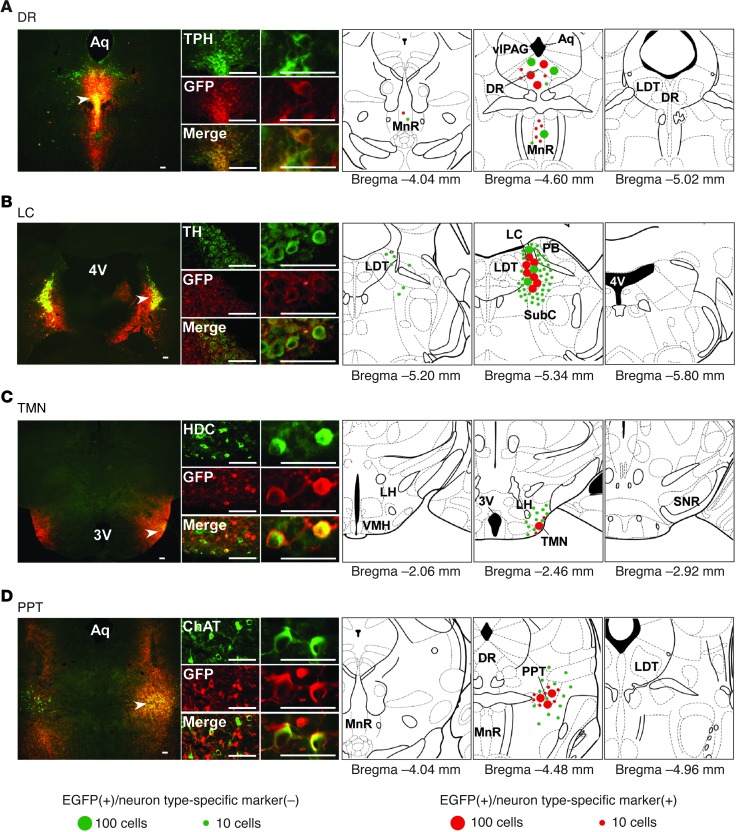
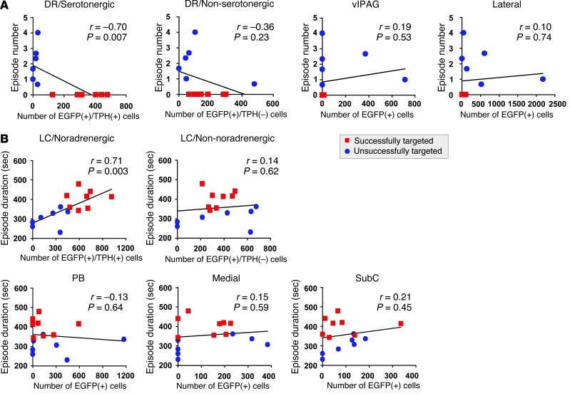
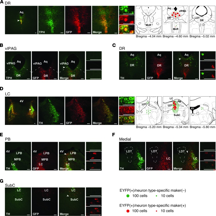
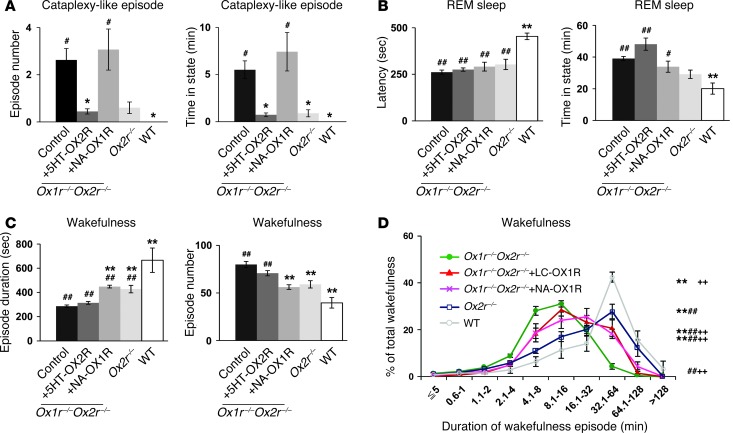
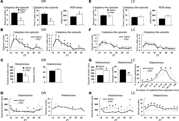
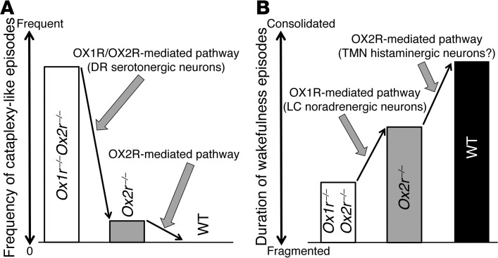
The loss of orexin neurons in humans is associated with the sleep disorder narcolepsy, which is characterized by excessive daytime sleepiness and cataplexy. Mice lacking orexin peptides, orexin neurons, or orexin receptors recapitulate human narcolepsy phenotypes, further highlighting a critical role for orexin signaling in the maintenance of wakefulness. Despite the known role of orexin neurons in narcolepsy, the precise neural mechanisms downstream of these neurons remain unknown. We found that targeted restoration of orexin receptor expression in the dorsal raphe (DR) and in the locus coeruleus (LC) of mice lacking orexin receptors inhibited cataplexy-like episodes and pathological fragmentation of wakefulness (i.e., sleepiness), respectively. The suppression of cataplexy-like episodes correlated with the number of serotonergic neurons restored with orexin receptor expression in the DR, while the consolidation of fragmented wakefulness correlated with the number of noradrenergic neurons restored in the LC. Furthermore, pharmacogenetic activation of these neurons using designer receptor exclusively activated by designer drug (DREADD) technology ameliorated narcolepsy in mice lacking orexin neurons. These results suggest that DR serotonergic and LC noradrenergic neurons play differential roles in orexin neuron-dependent regulation of sleep/wakefulness and highlight a pharmacogenetic approach for the amelioration of narcolepsy.


Figures

Figure 1
Figure 1. Region-specific restoration of orexin receptor expression in Ox1r–/–Ox2r–/– mice.
Coronal brain sections prepared from Ox1r–/–Ox2r–/– mice with targeted injection of AAV-EF1α/OX1R::EGFP (B and D) or AAV-EF1α/OX2R::EGFP (A and C) were double-stained with anti-GFP (red) and the neuronal type–specific marker (green) antibodies TPH (A), TH (B), HDC (C), and ChAT (D). Regions denoted by white arrowheads are shown at higher magnification. Schematics show the spread of OX1R::EGFP or OX2R::EGFP expression. Mean numbers of EGFP+ cells are shown by green or red circles, indicative of the absence or presence, respectively, of the neuronal type–specific marker. For bilaterally injected areas, only 1 side was shown. 3v, third ventricle; 4v, fourth ventricle, Aq, aqueduct; SNR, substantia nigra pars reticulata; VMH, ventromedial hypothalamus. Scale bars: 100 μm.
Figure 2
Figure 2. Restoration of orexin receptor expression in the DR and LC inhibits cataplexy-like episodes and consolidates wakefulness, respectively.
Control Ox1r–/–Ox2r–/– (n = 16), Ox1r–/–Ox2r–/–+DR-OX2R (n = 7), Ox1r–/–Ox2r–/–+LC-OX1R (n = 8), Ox1r–/–Ox2r–/–+TMN-OX2R (n = 4), Ox1r–/–Ox2r–/–+PPT-OX1R (n = 4), and WT (n = 4) mice were recorded. Mean values of 3 nights’ recordings are shown. Ox1r–/–Ox2r–/– controls were injected with AAV-CAG/EGFP into the DR or LC, AAV-Pet1/ChR2::EYFP into the DR, or AAV-PRSx8/ChR2::EYFP into the LC. (A) Number of and time spent in cataplexy-like episodes. (B) REM sleep latency and time spent in REM sleep. (C) Duration and number of wakefulness episodes during the dark phase. *P < 0.05, **P < 0.01 vs. Ox1r–/–Ox2r–/– control; #P < 0.05, ##P < 0.01 vs. WT. Values are mean ± SEM.
Figure 3
Figure 3. Suppression of narcoleptic symptoms is correlated with the number of serotonergic or noradrenergic neurons with orexin receptor in the DR or LC.
(A) Correlation between number of cataplexy-like episodes and number of EGFP+ cells in TPH+ DR, TPH DR, vlPAG, or the region lateral to the DR and MnR (including the paratrochlear nucleus and pontine reticular nucleus) of Ox1r–/–Ox2r–/– +DR-OX2R mice (Successfully targeted) and those with off-target restoration (Unsuccessfully targeted). (B) Correlation between wakefulness episode duration and number of EGFP+ cells in TH+ LC, TH LC, or regions lateral (including PB), medial, or ventral (including SubC) to the LC of Ox1r–/–Ox2r–/–+LC-OX1R mice and those with off-target restoration. Pearson r, P values, and regression lines are shown.
Figure 4
Figure 4. Restoration of orexin receptors in the DR or LC of Ox1r–/–Ox2r–/– mice using neuron type-selective promoters.
(AC) Coronal brain sections from Ox1r–/–Ox2r–/–+5HT-OX2R mice (with targeted injection of AAV-Pet1/OX2R::EYFP) were double-stained with anti-GFP antibody (red) and either anti-TPH (A and B) or anti-TH (C, for dopaminergic neurons in the DR) antibody (green). (DG) Sections from Ox1r–/–Ox2r–/–+NA-OX1R mice (with AAV-PRSx8/OX1R::EYFP injection) were double-stained with anti-GFP (red) and anti-TH (green) antibodies. Leaked expression of OX1R::EYFP in regions lateral (E, including PB), medial (F), and ventral (G, including SubC) to the LC (D) is shown. Regions denoted by white arrowheads are shown at higher magnification. Schematics show the spread of OX1R::EYFP or OX2R::EYFP expression. Mean numbers of EGFP+ cells are shown by green or red circles, indicative of the absence or presence, respectively, of the neuronal type–specific marker. For LC, only 1 side was shown. LPB, lateral PB; MPB, medial PB. Scale bars: 100 μm.
Figure 5
Figure 5. Restoration of orexin receptor expression selectively in DR serotonergic and LC noradrenergic neurons suppresses cataplexy-like episodes and ameliorates fragmentation of wakefulness, respectively.
(AC) Control virus–injected Ox1r–/–Ox2r–/– (as in Figure 2; n = 16), Ox1r–/–Ox2r–/–+5HT-OX2R (n = 6), Ox1r–/–Ox2r–/–+NA-OX1R (n = 5), Ox2r–/– (n = 5), and WT (n = 4) mice were recorded. Mean values of 3 nights’ recordings are shown. Data from Figure 2 are included for comparison. (A) Number of and time spent in cataplexy-like episodes. (B) REM sleep latency and time spent in REM sleep. (C) Duration and number of wakefulness episodes during the dark phase. *P < 0.05, **P < 0.01 vs. Ox1r–/–Ox2r–/– control; #P < 0.05, ##P < 0.01 vs. WT. (D) Time-weighted frequency histograms of wakefulness duration in control Ox1r–/–Ox2r–/– (n = 16), Ox1r–/–Ox2r–/–+LC-OX1R (n = 8), Ox1r–/–Ox2r–/–+NA-OX1R (n = 5), Ox2r–/– (n = 5), and WT (n = 4) mice, showing the wakefulness that occurred in episodes of each length as a percentage of total wakefulness in the dark phase. **P < 0.001 vs. Ox1r–/–Ox2r–/– control; ##P < 0.001 vs. WT; ++P < 0.001 vs. Ox2r–/–, 2-way repeated-measures ANOVA. Values are mean ± SEM.
Figure 6
Figure 6. Pharmacogenetic activation of DR serotonergic and LC noradrenergic neurons suppresses cataplexy-like episodes and consolidates wakefulness, respectively. orexin/ataxin-3 mice with DR serotonergic neuron–selective (AD; n = 7) or LC noradrenergic neuron–selective (EH; n = 8) expression of hM3Dq were injected with saline or CNO.
Mean values of 3 nights’ recordings are shown. (A and E) Number of and time spent in cataplexy-like episodes and REM sleep latency. (B and F) Hourly plots of number and total time of cataplexy-like episodes. (C and G) Duration and number of wakefulness episodes within 12 hours after saline or CNO administration at ZT 12 (arrows). (G) Time-weighted frequency histogram of wakefulness duration is also shown (P < 0.001, drug effect and interaction of drug and wakefulness duration, 2-way repeated-measures ANOVA). (D and H) Hourly plots of wakefulness duration and number within 12 hours after administration at ZT 12 (arrows). *P < 0.05, #P < 0.01, 2-tailed Student’s paired t test. Values are mean ± SEM.
Figure 7
Figure 7. Proposed model for OX1R- and OX2R-mediated pathways in suppressing narcoleptic symptoms.
(A) Prevention of cataplexy-like episodes. (B) Consolidation of wakefulness episodes.

References

    1. Sakurai T. The neural circuit of orexin (hypocretin): maintaining sleep and wakefulness. Nat Rev Neurosci. 2007;8(3):171–181. - PubMed
    1. Dauvilliers Y, Arnulf I, Mignot E. Narcolepsy with cataplexy. Lancet. 2007;369(9560):499–511. doi: 10.1016/S0140-6736(07)60237-2. - DOI - PubMed
    1. Chemelli RM, et al. Narcolepsy in orexin knockout mice: molecular genetics of sleep regulation. Cell. 1999;98(4):437–451. doi: 10.1016/S0092-8674(00)81973-X. - DOI - PubMed
    1. Hara J, et al. Genetic ablation of orexin neurons in mice results in narcolepsy, hypophagia, and obesity. Neuron. 2001;30(2):345–354. doi: 10.1016/S0896-6273(01)00293-8. - DOI - PubMed
    1. Hondo M, et al. Histamine-1 receptor is not required as a downstream effector of orexin-2 receptor in maintenance of basal sleep/wake states. Acta Physiol (Oxf). 2010;198(3):287–294. doi: 10.1111/j.1748-1716.2009.02032.x. - DOI - PMC - PubMed

Publication types

MeSH terms