. 2014 Jan;92(1):1-8.
doi: 10.1139/cjpp-2013-0063. Epub 2013 Aug 13.

Nonsurgically induced disuse muscle atrophy and neuromuscular dysfunction upregulates alpha7 acetylcholine receptors

Affiliations

Nonsurgically induced disuse muscle atrophy and neuromuscular dysfunction upregulates alpha7 acetylcholine receptors

Mohammed A S Khan et al. Can J Physiol Pharmacol. 2014 Jan.

Abstract

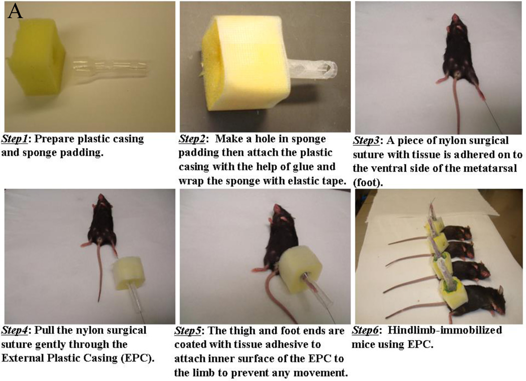
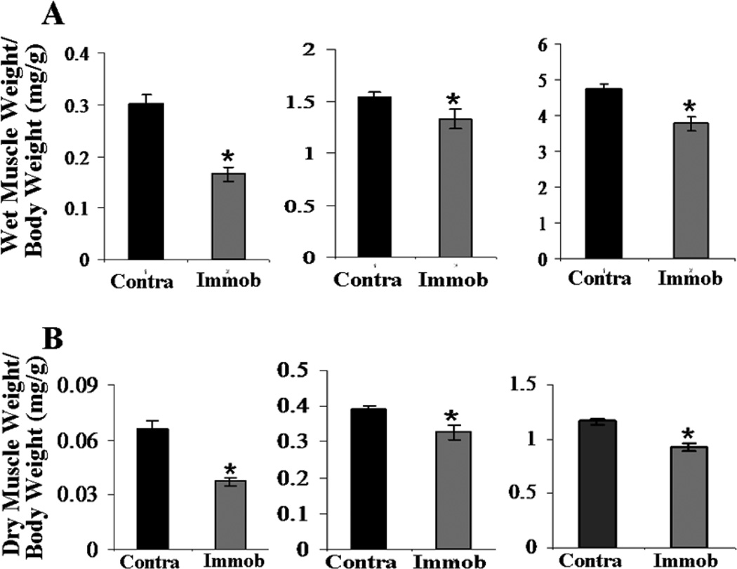
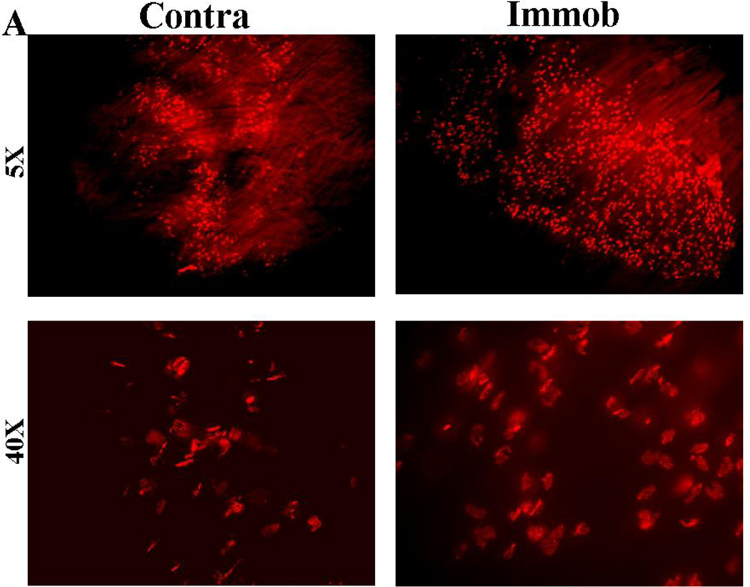
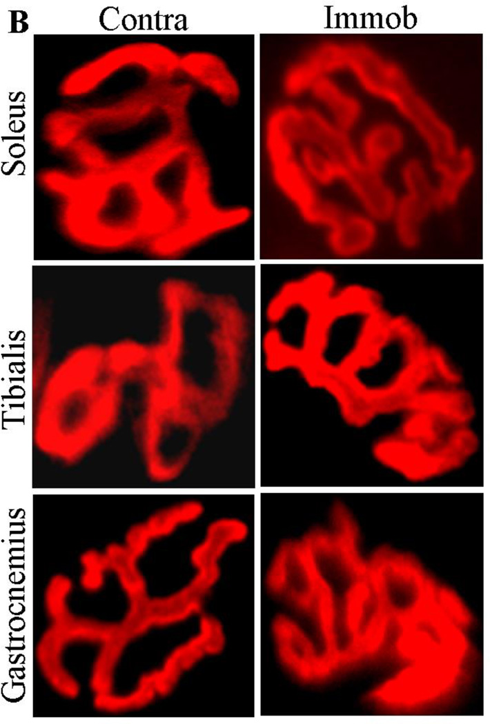
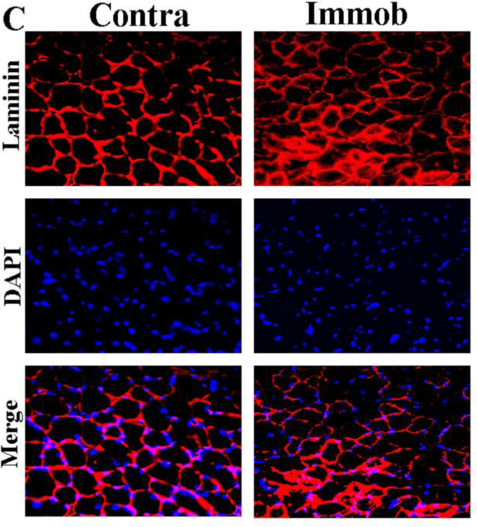
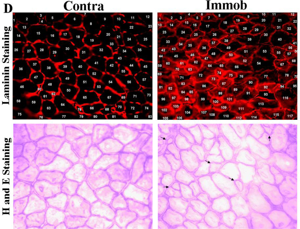
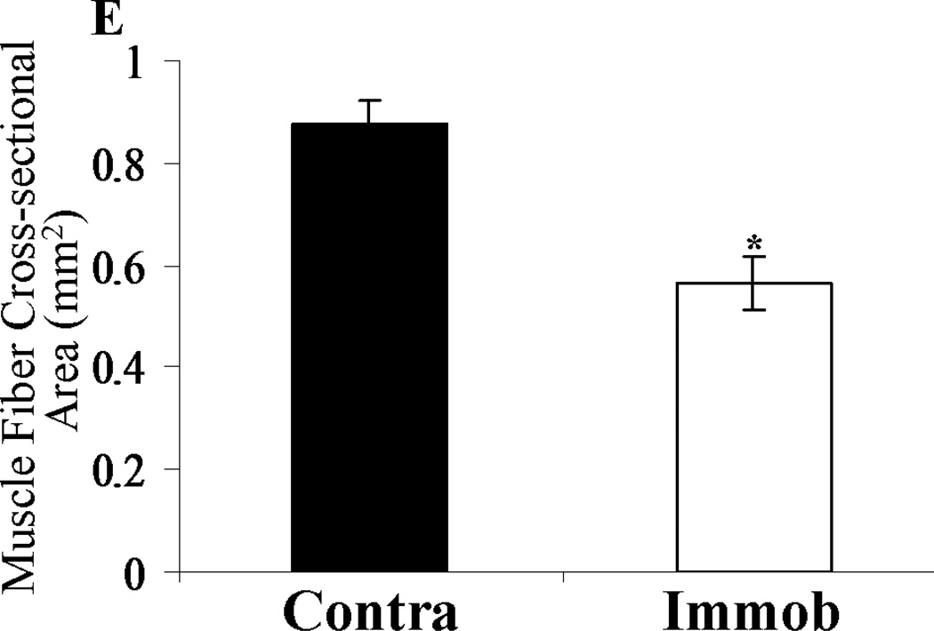
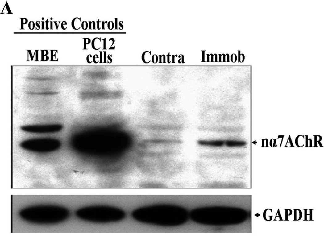
Previous models of muscle disuse have invariably used surgical methods that require the repetitive application of plaster casts. A method of disuse atrophy that does not require such repetitive applications is described herein. Modified plastic pipette tubing was applied to a single hindlimb (mouse), from thigh to foot, resulting in immobilization of the knee in the extension position, and the ankle in the plantar flexion position. This method resulted in the loss of soleus muscle to 11%, 22%, 39%, and 45% of its original mass at 3, 7, 14, and 21 days, respectively, in association with a significant decrease of tibialis twitch (25%) and tetanic tensions (26%) at 21 days, compared with the contralateral side and (or) sham-immobilized controls. Immunohistochemical analysis of the soleus using fluorescent α-bungarotoxin revealed a significant increase in the number of synapses per unit area (818 + 31 compared with 433 + 16/mm(2)) and an increase in muscle fibers per unit area (117 compared with 83/mm(2)), most likely related to the atrophy of muscle fibers bringing synapses closer. A 3-fold increase in alpha7 acetylcholine receptor (α7AChR) protein expression, along with increased expression of α1AChR subunit in the immobilized side compared with the contralateral side was observed. The physiology and pharmacology of the novel finding of upregulation of α7AChRs with disuse requires further study.


Figures

Fig. 1
Fig. 1. Stepwise process of hindlimb immobilization of mice by EPC
A. Brief description of each of 6 steps is described. B. Schematic, macroscopic and X-ray image of hindlimb immobilization of mouse. B1. Cartoon of EPC. B2. Picture of the hindlimb immobilized with EPC. B3. X-ray image of immobilized hindlimb and B4. Immobilized hindlimb after removal of EPC on day 14. The contralateral hindlimb is seen on the left.
Fig. 2
Fig. 2. Hindlimb immobilization by EPC and atrophy of muscles
A. Macroscopic views and muscle weights from immobilized (Immob) and contralateral (Contra) sides. Macroscopically, the muscles appeared smaller after immobilization. B. Microscopic analysis of immobilized (Immob) and contralateral (Contra) soleus muscles using Hematoxylin and Eosin staining. The cross-sectional area of whole soleus muscle measured by ImageJ indicated a reduction of ~50% after immobilization compared to contralateral side. Magnification 5X. C. Determination of soleus muscle weight showed the progressive loss of muscle mass on immobilized (Immob) side, at 3 (n=4, p >0.05), 7 (n=4, p <0.05), 14 (n=5, p <0.05) and 21 (n=4, p <0.05) days as compared to contralateral (Contra) side. The soleus muscle mass in sham-immobilized mice, not immobilized on the contralateral side, was used as control for 21 day immobilization group. The percentage of contralateral (Contra) side assumed as 100%. The muscle mass of soleus of contralateral side of immobilized and non-immobilized limb were not different. The data represent the mean ± standard error. Statistical analysis was done using one-way ANOVA with Tukey's multiple comparison tests. Asterisks represent p value (*p ≤0.05), as compared to contralateral side. NS represents not significant. Immobilization induced progressive loss of skeletal muscle mass.
Fig. 3
Fig. 3. Wet and dry weights of muscles and evoked muscle tensions
Wet (A) and dry (B) weights of soleus (left panel), tibialis (middle panel) and gastrocnemius (right panel) muscles. The muscle weights were normalized to body weight (mg/kg). C. Wet to dry weight ratio of soleus, tibialis and gastrocnemius muscles (n=5). The muscles did not show edematous tissues after immobilization. D. Nerve evoked single twitch and tetanic tensions were significantly reduced on the immobilized (Immob) side, shown as percentage of contralateral (Contra) side assumed as 100%. The data represent the mean ± standard error (n=5). Statistical analysis was done using Student t-test or one-way ANOVA with Tukey's multiple comparison tests. Asterisks represent p values (*p ≤ 0.05) as compared to contralateral side. EPC induced muscle weakness on immobilized side.
Fig. 4
Fig. 4. Muscle synapses and muscle fiber size
A. AChRs were stained using AlexaFlour 594 labeled-BTX in whole mount soleus muscle following immobilization for 21 days. The number of synapses per unit area on contralateral (Contra) and immobilized (Immob) sides counted at 5X magnification by five individuals indicated 433 + 16 and 818 + 31/mm2 synapses, respectively. Muscle synapses were increased due to atrophied muscle fibers on immobilized side compared to contralateral side. B. Morphological comparison of end-plates between immobilized (Immob) and contralateral (Contra) sides. The end-plates of soleus (top panel), tibialis (middle panel) and gastrocnemius (bottom panel) were stained using AlexaFlour 594 labeled-BTX. Endplates were more convoluted on immobilized side compared to contralateral side. C. Cross-sectional area of muscle fibers on immobilized (Immob) and contralateral (Contra) sides. The muscle membrane was stained using laminin specific antibody (top panel), which defined the margins of the muscle fiber, and the nuclei in the muscle fibers were localized with DAPI staining (middle panel) respectively, with merge image (bottom panel). Size of muscle fiber was reduced on immobilized side compared to contralateral side. D. Comparison of number of muscle fibers on contralateral side, Contra, (left panel) and immobilized side, Immob, (right panel). Laminin staining (upper panel). Each number represents one muscle fiber. The number of muscle fibers per unit area on contralateral (Contra) and immobilized (Immob) sides were 83 and 117/mm2, respectively. The higher number of muscle fibers per unit area indicates the atrophic muscle compared to contralateral side. H and E staining (lower panel). Arrows indicate the atrophied muscle fibers. Number of muscle fibers were decreased per unit area on immobilized side compared to contralateral side. E. Measurement of average cross-sectional area of soleus muscle fibers on contralateral side, Contra, (left panel) and immobilized side, Immob, (right panel) using ImageJ software. The average cross-sectional area of muscle fibers on contralateral (Contra) and immobilized (Immob) sides were 0.88 + 0.04 and 0.57 + 0.04 mm2, respectively. The data represent the mean ± standard error. Total 80 randomnly selected muscle fibers were counted from each side. Statistical analysis was done using Student t-test. Asterisks represent p values (*p < 0.05) as compared to contralateral side. Average muscle fiber cross-sectional area was reduced on immobilized side compared to contralateral side.
Fig. 5
Fig. 5. Expression of α7AChRs in soleus muscle
A. Western blot analysis using α7AChR specific antibody revealed increased expression of α7AChRs in soleus muscles. Mouse brain extract (MBE) and PC12 cells were used as positive control for α7AChR. Contralateral (Contra) and immobilized (Immob) sides at 21 days of immobilization. B. Densitometric analysis of α7AChRs. The data represent the mean ± standard error (n=3). Statistical analysis was done using Student t-test. Asterisks represent p values (*p ≤ 0.05) as compared to contralateral side. C. Western blot analysis of α1AChR subunit in soleus muscles. Contralateral (Contra) and immobilized (Immob) sides at 21 days of immobilization. Immobilization increased expression of α1- and α7-AChR subunits.

References

    1. Bajusz E. "Red" skeletal muscle fibers: relative independence of neural control. Science. 1964;45:938–939. - PubMed
    1. Brunelli S, Rovere-Querini P. The immune system and the repair of skeletal muscle. Pharmacol. Res. 2008;58:117–121. - PubMed
    1. Caron AZ, Drouin G, Desrosiers J, Trensz F, Grenier GA. Novel hindlimb immobilization procedure for studying skeletal muscle atrophy and recovery in mouse. J. Appl. Physiol. 2009;106:2049–2059. - PubMed
    1. Cartee GD, Bohn EE, Gibson BT, Farrar RP. Growth hormone supplementation increases skeletal muscle mass of old male fischer 344/brown norway rats. J. Gereontol.: Biol. Sci. 1996;51A:B214–B219. - PubMed
    1. Damrongrungruang T, Kuroda S, Kondo H, Aoki K, Ohya K, Kasugai S. A simple murine model for immobilization osteopenia. Clin. Orthop. Relat. Res. 2004;425:244–251. - PubMed

Publication types

MeSH terms

Substances

LinkOut - more resources