

Pulmonary emphysema subtypes on computed tomography: the MESA COPD study
- PMID: 24384106
- PMCID: PMC3882898
- DOI: 10.1016/j.amjmed.2013.09.020
Pulmonary emphysema subtypes on computed tomography: the MESA COPD study
Abstract
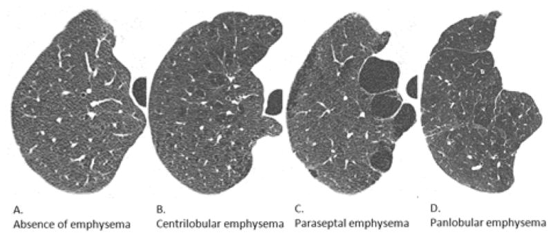
Background: Pulmonary emphysema is divided into 3 major subtypes at autopsy: centrilobular, paraseptal, and panlobular emphysema. These subtypes can be defined by visual assessment on computed tomography (CT); however, clinical characteristics of emphysema subtypes on CT are not well defined. We developed a reliable approach to visual assessment of emphysema subtypes on CT and examined if emphysema subtypes have distinct characteristics.
Methods: The Multi-Ethnic Study of Atherosclerosis COPD Study recruited smokers with chronic obstructive pulmonary disease (COPD) and controls ages 50-79 years with ≥ 10 pack-years. Participants underwent CT following a standardized protocol. Definitions of centrilobular, paraseptal, and panlobular emphysema were obtained by literature review. Six-minute walk distance and pulmonary function were performed following guidelines.
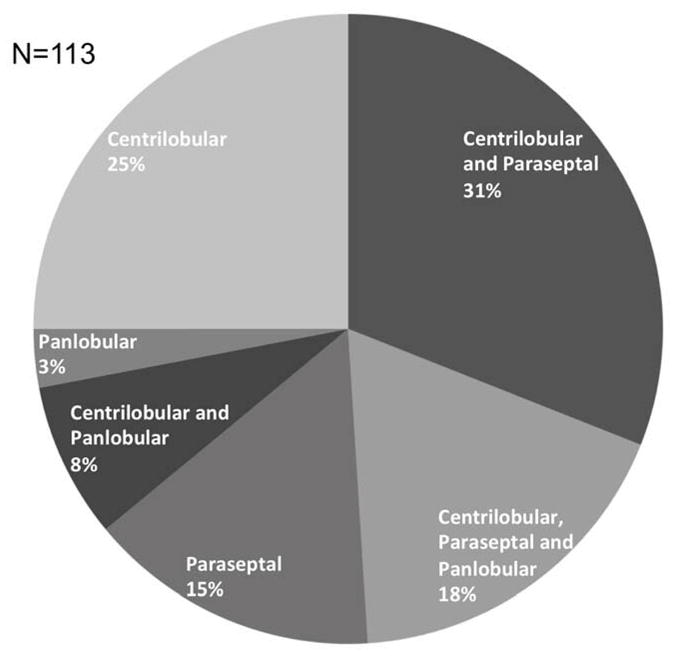
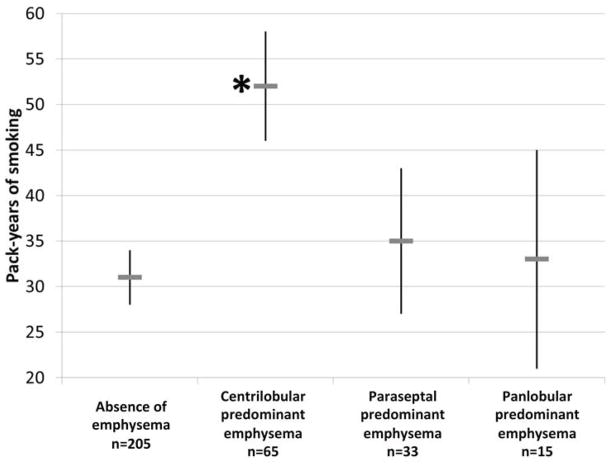
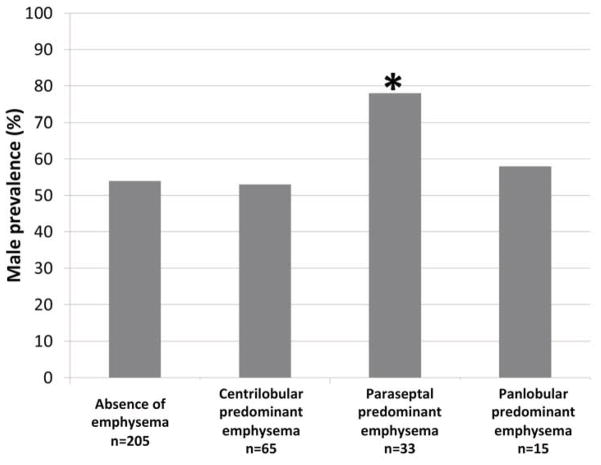
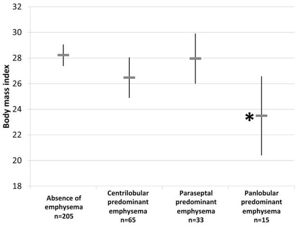
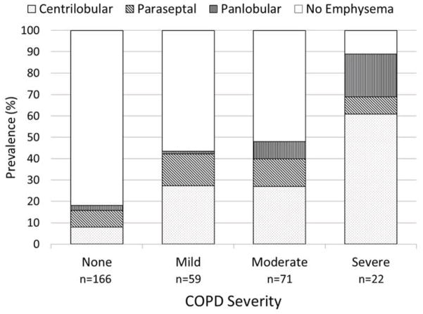
Results: Twenty-seven percent of 318 smokers had emphysema on CT. Interrater reliability of emphysema subtype was substantial (K: 0.70). Compared with participants without emphysema, individuals with centrilobular or panlobular emphysema had greater dyspnea, reduced walk distance, greater hyperinflation, and lower diffusing capacity. In contrast, individuals with paraseptal emphysema were similar to controls, except for male predominance. Centrilobular, but not panlobular or paraseptal, emphysema was associated with greater smoking history (+21 pack-years P <.001). Panlobular, but not other types of emphysema, was associated with reduced body mass index (-5 kg/m(2); P = .01). Other than for dyspnea, these findings were independent of the forced expiratory volume in 1 second. Seventeen percent of smokers without COPD on spirometry had emphysema, which was independently associated with reduced walk distance.
Conclusions: Emphysema subtypes on CT are common in smokers with and without COPD. Centrilobular and panlobular emphysema, but not paraseptal emphysema, have considerable symptomatic and physiological consequences.
Keywords: Centrilobular; Computed tomography; Emphysema; Panlobular; Paraseptal.
Copyright © 2014 Elsevier Inc. All rights reserved.
Conflict of interest statement
Figures
References
-
- Hoyert DL, Xu J. Deaths: Preliminary Data for 2011. National vital statistics reports: from the Centers for Disease Control and Prevention, National Center for Health Statistics. National Vital Statistics System. 2012;61:1–52. - PubMed
-
- Celli BR, MacNee W, Force AET. Standards for the diagnosis and treatment of patients with COPD: a summary of the ATS/ERS position paper. Eur Respir J. 2004;23:932–46. - PubMed
-
- The definition of emphysema. Report of a National Heart, Lung, and Blood Institute, Division of Lung Diseases workshop. Am Rev Respir Dis. 1985;132:182–5. - PubMed
-
- Hayhurst MD, MacNee W, Flenley DC, et al. Diagnosis of pulmonary emphysema by computerised tomography. Lancet. 1984;2:320–2. - PubMed
-
- Bergin C, Müller N, Nichols DM, et al. The diagnosis of emphysema. A computed tomographic-pathologic correlation. Am Rev Respir Dis. 1986;133:541–6. - PubMed
Publication types
MeSH terms
Grants and funding
LinkOut - more resources
Full Text Sources
Other Literature Sources
Medical
Molecular Biology Databases
