. 2014 Feb 15;386(2):302-7.
doi: 10.1016/j.ydbio.2013.12.035. Epub 2013 Dec 31.

Oral-aboral axis specification in the sea urchin embryo, IV: hypoxia radializes embryos by preventing the initial spatialization of nodal activity

Affiliations

Oral-aboral axis specification in the sea urchin embryo, IV: hypoxia radializes embryos by preventing the initial spatialization of nodal activity

James A Coffman et al. Dev Biol. .

Abstract

The oral-aboral axis of the sea urchin embryo is specified conditionally via a regulated feedback circuit involving the signaling gene nodal and its antagonist lefty. In normal development nodal activity becomes localized to the prospective oral side of the blastula stage embryo, a process that requires lefty. In embryos of Strongylocentrotus purpuratus, a redox gradient established by asymmetrically distributed mitochondria provides an initial spatial input that positions the localized domain of nodal expression. This expression is perturbed by hypoxia, leading to development of radialized embryos lacking an oral-aboral axis. Here we show that this radialization is not caused by a failure to express nodal, but rather by a failure to localize nodal activity to one side of the embryo. This occurs even when embryos are removed from hypoxia at late cleavage stage when nodal is first expressed, indicating that the effect involves the initiation phase of nodal activity, rather than its positive feedback-driven amplification and maintenance. Quantitative fluorescence microscopy of MitoTracker Orange-labeled embryos expressing nodal-GFP reporter gene revealed that hypoxia abolishes the spatial correlation between mitochondrial distribution and nodal expression, suggesting that hypoxia eliminates the initial spatial bias in nodal activity normally established by the redox gradient. We propose that absent this bias, the initiation phase of nodal expression is spatially uniform, such that the ensuing Nodal-mediated community effect is not localized, and hence refractory to Lefty-mediated enforcement of localization.

Keywords: Axis; Embryo; Hypoxia; Mitochondria; Nodal; Polarity.


Figures

Figure 1
Figure 1
Effects of timed embryo exposures to hypoxia on Sp-nodal expression and oral-aboral axis development. (A) Relative per-embryo levels of nodal transcripts at 9, 12, and 18 hours post-fertilization in normoxically and hypoxically cultured embryos. Error bars are standard deviation of technical replicates. (B) Spatial expression of nodal, revealed by confocally imaged fluorescent WMISH at 12 hpf in normoxically and hypoxically cultured embryos from the experiment shown in (A). (C) Early temporal expression profiles of nodal in normoxically and hypoxically cultured embryos, assayed as in (A), but in a different year with a different batch of embryos. (D) Relative per-embryo levels of nodal transcripts at 16 hpf in embryos cultured hypoxically up to 8 or 16 hpf, compared to normoxic controls, from the experiment shown in (C). (E) Lateral view showing the radialized phenotype of a 2 day (55 hr) embryo cultured hypoxically for 9 hrs beginning at the 2-cell stage. (F) Quantification of prism stage oral-aboral axial phenotypes following different durations of hypoxic culture (from the time-course experiment shown in Fig. 1C). For each sample 50 embryos were counted and scored as either morphologically normal (bilaterally symmetric), intermediate, or radialized.
Figure 2
Figure 2
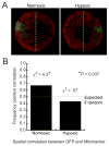
Effects of hypoxia on spatial correlation between nodal-5P-GFP expression and mitochondrial distribution. (A) Examples of normoxically- and hypoxically-cultured 12 hr embryos labelled with MitoTracker Orange (red), expressing microinjected nodal-5P-GFP (green). The dotted line illustrates how the images were divided into GFP expressing and non-expressing halves for quantifying average MitoTracker pixel intensity, as described previously (Coffman et al., 2009). In the normoxically-cultured embryo shown, the expressing half has a higher than average MitoTracker signal, whereas in the hypoxically-cultured embryo the expressing half has a lower than average MitoTracker signal. (B) Correlation between nodal-5P-GFP expression and a higher than average MitoTracker signal in the expressing half, in sibling embryos cultured normoxically and hypoxically. A Chi-squared test with one degree of freedom was used to assess statistical significance of the difference between the observed correlation and what would be expected at random. For the normoxic embryos n = 39; for the hypoxic embryos n = 28.
Figure 3
Figure 3
Knockdown of nodal in embryo halves rescues a secondary axis and entrains oral-aboral polarity in hypoxically-cultured embryos. (A) Diagram of experimental setup. (B) Quantification of axial phenotypes obtained at 3 days post fertilization (dpf) after the embryos were cultured hypoxically for 9 hrs following injection, with images of representative examples. Note that in some initial experiments (not shown) the hypoxically-cultured sham controls (including uninjected controls subjected to all steps in the injection procedure except for the injection itself) unaccountably developed a secondary axis. In those experiments the hypoxically-cultured non-sham embryos were nevertheless radialized. (C) Quantification of the ultimate location of the fluorescent lineage tracer, with example images of injected embryos at 3 dpf showing the oral and aboral patterns. The numbers for the control embryos were obtained from experiments in which the controls were not radialized.
Figure 4
Figure 4
Updated model of oral-aboral axis specification in the sea urchin embryo that accounts for the radializing effect of hypoxia. (A) Diagram showing the spatial relationship between mitochondrial distribution in the cleavage stage embryo (blue); nodal mRNA expression (and by 12 hpf, lefty as well) at cleavage and blastula stage (green); Lefty protein diffusion and resultant inhibition of Nodal signaling at blastula stage (red), and the oral-aboral axis of the pluteus. (B) Diagram relating the redox gradient in the cleavage stage embryo to the spatially differential rates of nodal mRNA accumulation, leading to a localized (pre-oral) community effect regulatory state that activates lefty, whose extracellular product enforces that localization. (C) Diagram depicting the model suggested by the findings presented in this paper: hypoxia abolishes the redox gradient that normally generates the initial spatial bias in nodal mRNA accumulation, allowing development of a non-localized Nodal-mediated community effect within presumptive ectoderm.

References

    1. Agca C, Klein WH, Venuti JM. Reduced O2 and elevated ROS in sea urchin embryos leads to defects in ectoderm differentiation. Dev Dyn. 2009;238:1777–1787. - PubMed
    1. Ben-Tabou de-Leon S, Su YH, Lin KT, Li E, Davidson EH. Gene regulatory control in the sea urchin aboral ectoderm: spatial initiation, signaling inputs, and cell fate lockdown. Dev Biol. 2013;374:245–54. - PMC - PubMed
    1. Bolouri H, Davidson EH. The gene regulatory network basis of the “community effect,” and analysis of a sea urchin embryo example. Dev Biol. 2010;340:170–8. - PMC - PubMed
    1. Bradham CA, Oikonomou C, Kuhn A, Core AB, Modell JW, McClay DR, Poustka AJ. Chordin is required for neural but not axial development in sea urchin embryos. Dev Biol. 2009;328:221–33. - PMC - PubMed
    1. Chen Y, Schier AF. Lefty proteins are long-range inhibitors of squint-mediated nodal signaling. Curr Biol. 2002;12:2124–8. - PubMed

Publication types

MeSH terms

LinkOut - more resources