. 2014 Jan 1;6(1):a008995.
doi: 10.1101/cshperspect.a008995.

Regulation of receptor tyrosine kinase ligand processing

Affiliations

Regulation of receptor tyrosine kinase ligand processing

Colin Adrain et al. Cold Spring Harb Perspect Biol. .

Abstract

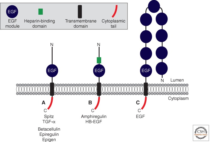
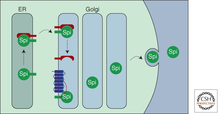
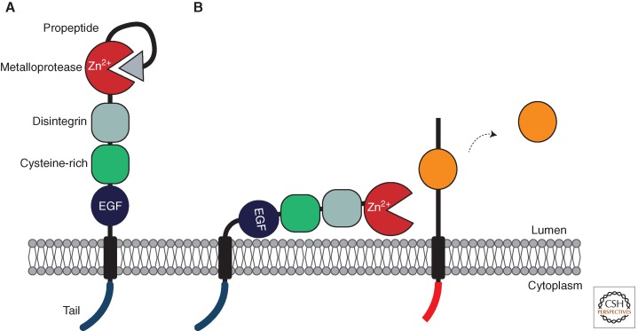
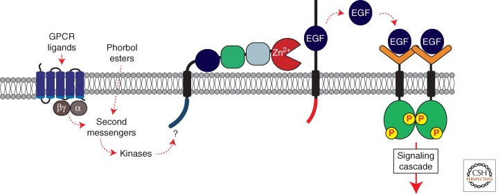
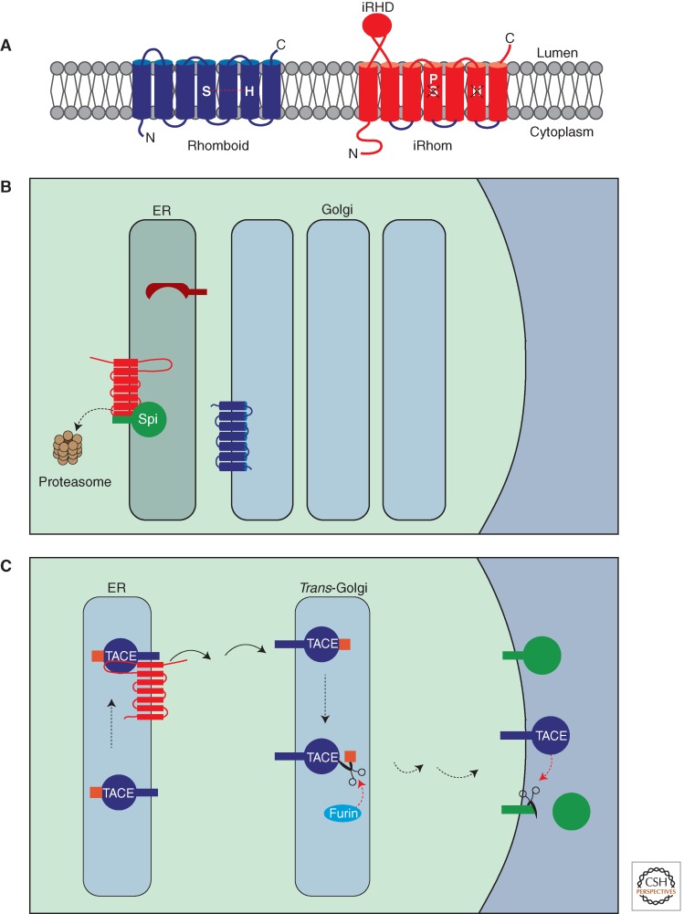
A primary mode of regulating receptor tyrosine kinase (RTK) signaling is to control access of ligand to its receptor. Many RTK ligands are synthesized as transmembrane proteins. Frequently, the active ligand must be released from the membrane by proteolysis before signaling can occur. Here, we discuss RTK ligand shedding and describe the proteases that catalyze it in flies and mammals. We focus principally on the control of EGF receptor ligand shedding, but also refer to ligands of other RTKs. Two prominent themes emerge. First, control by regulated trafficking and cellular compartmentalization of the proteases and their ligand substrates plays a key role in shedding. Second, many external signals converge on the shedding proteases and their control machinery. Proteases therefore act as regulatory hubs that integrate information that the cell receives and translate it into precise outgoing signals. The activation of signaling by proteases is therefore an essential element of the cellular communication machinery.


Figures

Figure 1.
Figure 1.
Topology of epidermal growth factor receptor (EGFR) ligands. EGFR ligands are type I transmembrane proteins with an extracellular (luminal) amino-terminus and a cytoplasmic carboxyl terminus. The domain structure of various EGFR ligands is indicated. (A) Drosophila Spitz, and the mammalian EGFR ligands TGF-α, Betacellulin, Epiregulin, and Epigen have a basic structure containing an amino-terminal prodomain and a bioactive EGF domain (indicated in blue). (B) Amphiregulin and HB-EGF contain a heparin binding motif amino terminal to the EGF domain (indicated in green); this facilitates binding to extracellular proteoglycans. Proteolytic cleavage occurs within the juxtamembrane domain between the EGF domain and the TMD; proteolytic removal of the amino-terminal prodomain also occurs (A,B). (C) Epidermal growth factor (EGF) contains additional EGF domains. The EGF domain closest to the membrane can activate the EGFR, whereas the remaining eight EGF domains cannot. The role of these is unclear, although they may play a role in regulating cell–cell adhesion. Cleavage liberates the bioactive EGF domain from the transmembrane precursor; depending on the tissue/context, the other EGF modules may either remain on the soluble molecule, or are cleaved off.
Figure 2.
Figure 2.
Domain structure of a rhomboid protease. Secretase rhomboids are polytopic transmembrane proteins with a cytoplasmic amino terminus and six or seven transmembrane domains. The catalytic serine and histidine residues are positioned within the upper third of transmembrane helices 4 and 6, respectively.
Figure 3.
Figure 3.
Regulated Spitz trafficking controls EGFR activation in Drosophila. Spitz is synthesized in the endoplasmic reticulum (ER) as a transmembrane precursor. Exit of Spitz from the ER to the Golgi requires the chaperone protein, Star (illustrated in red). On entry to the Golgi, Spitz encounters rhomboid (illustrated in blue) and undergoes proteolysis within the transmembrane domain. Spitz can now be secreted, thereby facilitating EGFR activation on a nearby cell.
Figure 4.
Figure 4.
Domain structure and activation of ADAM metalloproteases. ADAMs are type I transmembrane proteins containing extracellular (luminal) amino termini and cytoplasmic carboxyl termini. (A) ADAMs are synthesized as a zymogen proform that lacks proteolytic activity, because the prodomain binds within the active site cleft. Removal of the prodomain by autocatalysis or by the proportein convertase, furin, is required before the enzyme can be active. (B) After processing, the prodomain may remain bound to the active site, and may require displacement before the ADAM can be active. When activated by signals, ADAMs cleave their substrates with a region just outside the membrane (within the juxtamembrane region).
Figure 5.
Figure 5.
Transctivation of the EGFR by G protein-coupled receptors (GPCRs). Activation of GPCRs by agonists triggers a signaling cascade involving second messengers including Ca2+ and protein kinase C (PKC). This induces TACE cleavage of EGFR ligands (including HB-EGF), culminating in EGFR activation. How these signals trigger TACE activation remains unclear (see text). Phorbol esters such as PMA can also trigger TACE via a mechanism that involves PKC.
Figure 6.
Figure 6.
Regulation of RTK signaling by iRhoms. (A) Comparison of an active rhomboid and an iRhom. In comparison with an active rhomboid (left), iRhoms (right) contain an extended cytoplasmic amino terminus and a globular cysteine-rich domain called the iRhom homology domain (iRHD) within the lumen of the ER. All iRhoms have a conserved proline residue immediately amino terminal to the serine in TMD 4; this renders iRhoms proteolytically inactive. (B) Drosophila iRhom regulates ER-associated degradation of EGFR ligands. Spitz is normally trafficked out of the ER by Star and encounters rhomboid in the Golgi (Fig. 3). However in the presence of iRhom, Spitz is retained in the ER and instead, shunted into the ER-associated degradation (ERAD). This results in its dislocation from the ER membrane and degradation by the proteasome. As a result, no Spitz enters the Golgi for cleavage and EGFR signaling is attenuated. (C) Regulation of TACE trafficking by mammalian iRhom2. TACE is synthesized in the ER as an inactive zymogen containing the prodomain (the TACE prodomain is indicated in orange). iRhom is required for trafficking of TACE into the Golgi, where it undergoes prodomain cleavage by furin. Active TACE can then cleave its substrates in the late Golgi or on the cell surface.

References

    1. Adrain C, Freeman M 2012. New lives for old: Evolution of pseudoenzyme function illustrated by iRhoms. Nat Rev Mol Cell Biol 13: 489–498 - PubMed
    1. Adrain C, Strisovsky K, Zettl M, Hu L, Lemberg MK, Freeman M 2011. Mammalian EGF receptor activation by the rhomboid protease RHBDL2. EMBO Rep 12: 421–427 - PMC - PubMed
    1. Adrain C, Zettl M, Christova Y, Taylor N, Freeman M 2012. Tumor necrosis factor signaling requires iRhom2 to promote trafficking and activation of TACE. Science 335: 225–228 - PMC - PubMed
    1. Arduise C, Abache T, Li L, Billard M, Chabanon A, Ludwig A, Mauduit P, Boucheix C, Rubinstein E, Le Naour F 2008. Tetraspanins regulate ADAM10-mediated cleavage of TNF-α and epidermal growth factor. J Immunol 181: 7002–7013 - PubMed
    1. Asakura M, Kitakaze M, Takashima S, Liao Y, Ishikura F, Yoshinaka T, Ohmoto H, Node K, Yoshino K, Ishiguro H, et al. 2002. Cardiac hypertrophy is inhibited by antagonism of ADAM12 processing of HB-EGF: Metalloproteinase inhibitors as a new therapy. Nat Med 8: 35–40 - PubMed

Publication types

LinkOut - more resources