

Review
doi: 10.1038/nrmicro3160.
Epub 2014 Jan 2.
Bacteria-autophagy interplay: a battle for survival
Affiliations
- PMID: 24384599
- PMCID: PMC7097477
- DOI: 10.1038/nrmicro3160
Item in Clipboard
Review
Bacteria-autophagy interplay: a battle for survival
Nat Rev Microbiol.
2014 Feb.
Abstract
Autophagy is a cellular process that targets proteins, lipids and organelles to lysosomes for degradation, but it has also been shown to combat infection with various pathogenic bacteria. In turn, bacteria have developed diverse strategies to avoid autophagy by interfering with autophagy signalling or the autophagy machinery and, in some cases, they even exploit autophagy for their growth. In this Review, we discuss canonical and non-canonical autophagy pathways and our current knowledge of antibacterial autophagy, with a focus on the interplay between bacterial factors and autophagy components.
Conflict of interest statement
The authors declare no competing financial interests.
Figures
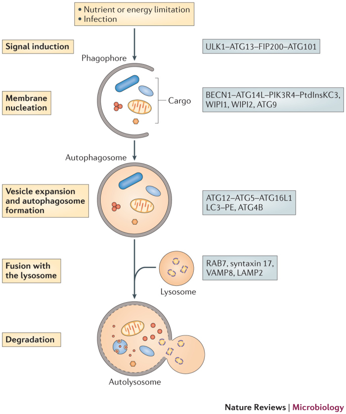
On stimulation of autophagy, a small membrane sac, known as the phagophore, is assembled and starts to elongate to enclose cytoplasmic components. The phagophore expands and grows into a double-membrane compartment, known as the autophagosome, which sequesters cytoplasmic targets, such as proteins, organelles and microorganisms. The autophagosome fuses with the lysosome to generate the autolysosome, in which the cargo is degraded by hydrolytic enzymes. Key proteins involved in autophagy in mammals are shown on the right. ATG, autophagy-related; ATG14L, ATG14-like; ATG16L1, ATG16-like 1; BECN1, beclin 1; FIP200, FAK family kinase-interacting protein of 200 kDa; LAMP2, lysosomal-associated membrane glycoprotein 2; LC3, microtubule-associated protein 1 light chain 3; PE, phosphatidylethanolamine; PIK3R4, phosphoinositide 3-kinase regulatory subunit 4; PtdInsKC3, phosphatidylinositol-3 kinase class III; ULK1, Unc-51-like kinase; VAMP8, vesicle-associated membrane protein 8; WIPI, WD-repeat domain phosphoinositide-interacting. PowerPoint slide
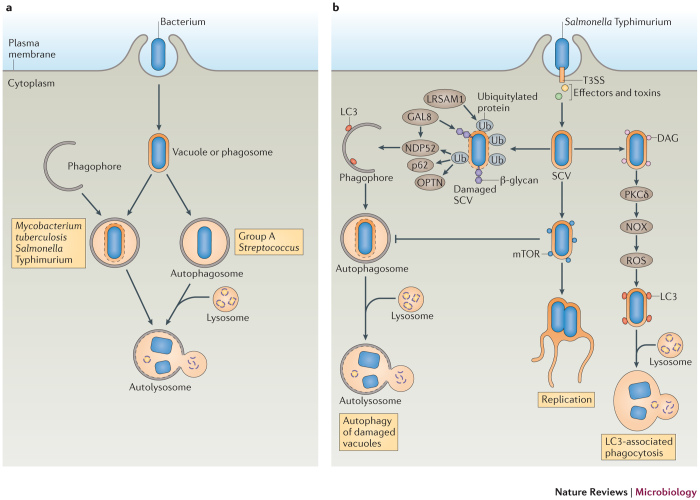
a | Schematic diagram of the ways that autophagy targets intracellular bacteria. After invasion of host cells, the bacterium resides in a bacterium-containing vacuole (or phagosome). Some bacteria damage the vacuolar or phagosomal membrane and eventually escape into the cytosol. Autophagy can target bacteria in damaged vacuoles or phagosomes (as with, for example, Mycobacterium tuberculosis and Salmonella enterica subsp. enterica serovar Typhimurium) or in the cytosol (as with, for example, Group A Streptococcus) and deliver them to the lysosome for degradation. b | Model of the antibacterial autophagy of S. Typhimurium. The bacterium resides in a Salmonella-containing vacuole (SCV) after invading epithelial cells. Early after infection (∼1 hour), the membrane of a subset of SCVs is damaged and the bacterium is exposed to the cytoplasm, where it becomes associated with ubiquitylated proteins in a process that depends on the E3 ubiquitin ligase leucine-rich repeat and sterile α-motif-containing 1 (LRSAM1). Next, adaptor proteins, including p62, NDP52 (nuclear dot protein 52 kDa) and optineurin (OPTN) are recruited to the SCV by binding to ubiquitin, and they recruit autophagosomes by interacting with microtubule-associated protein 1 light chain 3 (LC3), ultimately resulting in autolysosome formation. The damaged SCV membrane also exposes β-glycans to the cytoplasm and recruits galectin 8 (GAL8), which binds to NDP52 and further recruits LC3. Also at ∼1 hour post-infection, a subset of bacteria recruits diacylglycerol (DAG) to the undamaged SCVs. DAG activates protein kinase Cδ (PKCδ), which activates NADPH oxidase (NOX) and promotes reactive oxygen species (ROS) production, in turn inducing LC3-associated phagocytosis of bacteria. At ∼4 hours post-infection, mammalian target of rapamycin (mTOR) is redistributed to the SCV membrane, which inhibits autophagy induction, resulting in autophagy escape and enabling bacteria to replicate in SCVs. T3SS, type III secretion system. PowerPoint slide
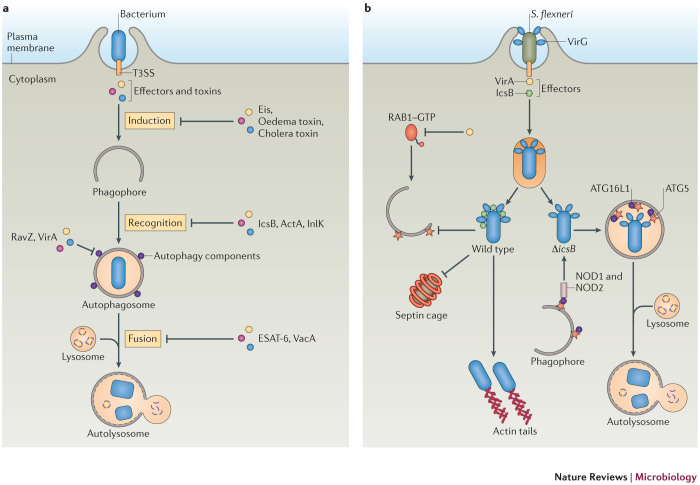
a | A diagram of how bacteria interfere with the autophagy machinery. Bacteria actively invade mammalian cells and secrete effectors to modulate the host cell machinery. Some bacteria use their effectors or toxins to interfere with the autophagy machinery at various stages in order to escape autophagic killing. Methods include inhibiting the autophagy induction signal (as with Eis (enhanced intracellular survival) from Mycobacterium tuberculosis, oedema factor toxin from Bacillus anthracis and cholera toxin from Vibrio cholerae), inhibiting recognition by the autophagy machinery (as with IcsB from Shigella flexneri, and ActA and internalin K (InlK) from Listeria monocytogenes), directly interfering with autophagy components (as with RavZ from Legionella pneumophila and VirA from S . flexneri) or blocking autophagosome fusion with the lysosome (as with ESAT-6 (early secreted antigenic target of 6 kDa) from M . tuberculosis and VacA from Helicobacter pylori). b | Model of S. flexneri evasion of autophagy. Wild-type bacteria escape the phagosomal compartment into the cytosol, where they recruit actin tails and replicate. The secreted effector IcsB binds to the bacterial surface protein VirG to block autophagy-related 5 (ATG5). IcsB also prevents formation of septin cages around the bacteria. The effector protein VirA inactivates RAB1 GTPase through its GTPase-activating protein (GAP) activity and inhibits autophagy. ΔicsB-mutant bacteria are recognized by nucleotide-binding oligomerization domain-containing 1 (NOD1) and NOD2, which interact with ATG16-like 1 (ATG16L1) and recruit autophagosomes to the invaded bacteria. In addition, ATG5 binds VirG and targets bacteria to autophagosomes. T3SS, type 3 secretion system. PowerPoint slide
References
Publication types
MeSH terms
LinkOut - more resources
Full Text Sources
Other Literature Sources
