. 2014 Feb 4;110(3):741-52.
doi: 10.1038/bjc.2013.793. Epub 2014 Jan 2.

Circulating galectins -2, -4 and -8 in cancer patients make important contributions to the increased circulation of several cytokines and chemokines that promote angiogenesis and metastasis

Affiliations

Circulating galectins -2, -4 and -8 in cancer patients make important contributions to the increased circulation of several cytokines and chemokines that promote angiogenesis and metastasis

C Chen et al. Br J Cancer. .

Abstract

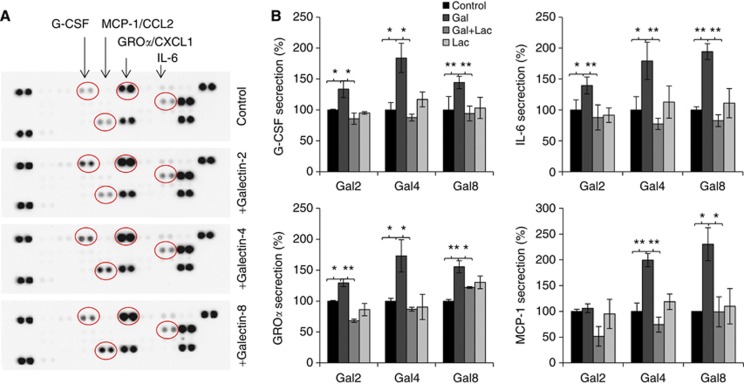
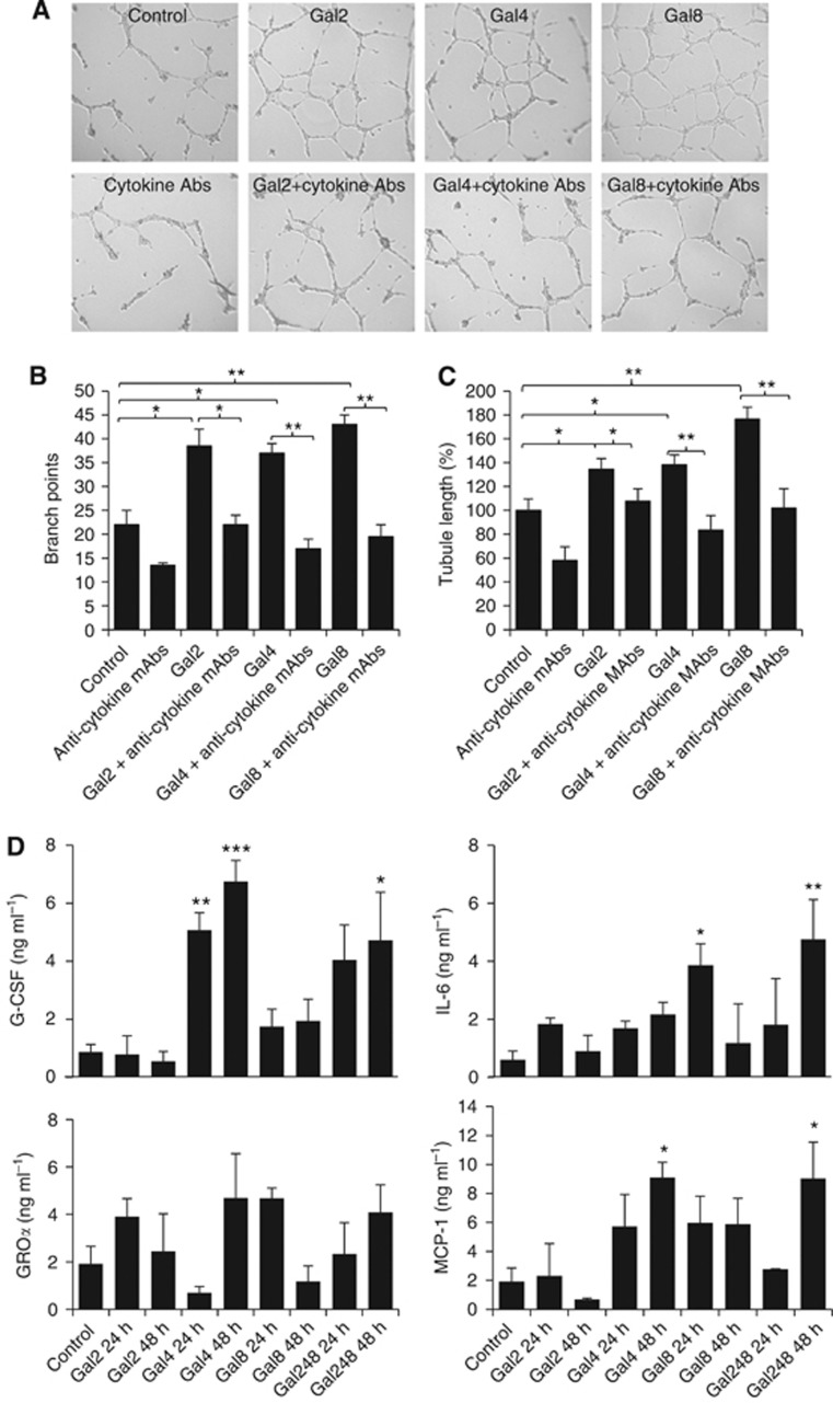
Background: Circulating concentrations of the cytokines interleukin-6 (IL-6), granulocyte colony-stimulating factor (G-CSF) and chemokines monocyte chemotatic protein 1 (MCP-1)/CCL2 and growth-regulator oncogene α (GROα)/chemokine C-X-C motif ligand 1 are commonly increased in cancer patients and they are increasingly recognised as important promoters, via divergent mechanisms, of cancer progression and metastasis.

Methods: The effect of galectins-2, -4 and -8, whose circulating levels are highly increased in cancer patients, on endothelial secretion of cytokines was assessed in vitro by cytokine array and in mice. The relationship between serum levels of galectins and cytokines was analysed in colon and breast cancer patients.

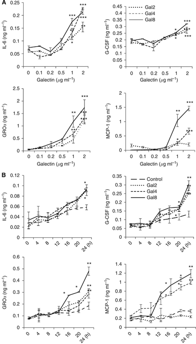
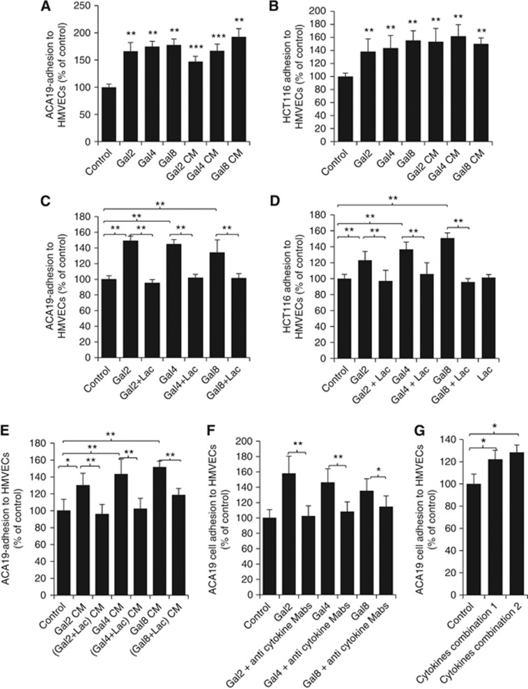
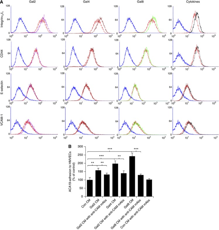
Results: Galectins-2, -4 and -8 at pathological concentrations induce secretion of G-CSF, IL-6, MCP-1 and GROα from the blood vascular endothelial cells in vitro and in mice. Multiple regression analysis indicates that increased circulation of these galectins accounts for 41∼83% of the variance of these cytokines in the sera of colon and breast cancer patients. The galectin-induced secretion of these cytokines/chemokines is shown to enhance the expression of endothelial cell surface adhesion molecules, causing increased cancer-endothelial adhesion and increased endothelial tubule formation.

Conclusion: The increased circulation of galectins -2, -4 and -8 in cancer patients contributes substantially to the increased circulation of G-CSF, IL-6 and MCP-1 by interaction with the blood vascular endothelium. These cytokines and chemokines in turn enhance endothelial cell activities in angiogenesis and metastasis.


Figures

Figure 1
Figure 1
Galectins -2, -4 and -8 induce cytokine secretion from endothelial cells. Human micro-vascular lung endothelial cells were incubated with 1.5 μg ml−1 galectins -2, -4, -8 or BSA for 24 h before the cytokine levels in the culture media were analysed by the cytokine array (A). In (B), HMVEC-Ls cells were treated with 1.5 μg ml−1 galectins -2, -4, -8 or BSA in the presence or absence of 200 μg ml−1 lactose for 24 h before the G-CSF, IL-6, GROα and MCP-1 levels in the cultured media were determined by ELISA. The data are expressed as percentage compared with BSA-treated controls from three independent experiments, each in triplicate. *P<0.05, **P<0.01 (ANOVA, Bonferroni).
Figure 2
Figure 2
Galectins -2, -4 and -8-induced endothelial secretions of cytokines are time- and dose-dependent. Human micro-vascular lung endothelial cells were treated with various concentrations of galectins -2, -4 or -8 for 24 h (A), or with 1.5 μg ml−1 galectins -2, -4, -8 or BSA for various times (B), before the G-CSF, IL-6, GROα and MCP-1 levels in the cultured media were determined. The data are expressed as mean±s.d. of triplicate determinations *P<0.05, **P<0.01, ***P<0.001 (ANOVA, Bonferroni).
Figure 3
Figure 3
Galectin-induced cytokine secretion enhances cancer cell-endothelial adhesion. (A and B) The presence of galectins -2, -4 or -8 increase cancer cell adhesion to HMVEC-Ls. Human micro-vascular lung endothelial cells were treated with 1.5 μg ml−1 galectins -2, -4 or -8 for 24 h. The cells were either washed and used for subsequent assessment of ACA19 (A) or HCT116 (B) cell adhesion, or the culture medium (CM) were collected and used for subsequent assessment of ACA19 (A) or HCT116 (B) cell adhesion. (C and D) The galectin-induced cancer cell adhesion is inhibited by lactose. Human micro-vascular lung endothelial cells were treated with 1.5 μg ml−1 galectins -2, -4 or -8 in the presence or absence of 200 μg ml−1 lactose for 24 h. The HMVEC-Ls were then used for assessment of ACA19 (C) and HCT116 (D) cell adhesion or the culture medium were collected and used for assessment of ACA19 cell adhesion to fresh HMVEC-Ls (E). (F) Galectin-mediated cancer cell-endothelial adhesion is inhibited by neutralising anti-cytokine antibodies. Human micro-vascular lung endothelial cells were treated with or without 1.5 μg ml−1 galectins -2, -4 or -8 in the presence or absence of antibodies against G-CSF (5 ng ml−1), IL-6 (3 ng ml−1), GROα (20 ng ml−1) and MCP-1(20 ng ml−1) in combination for 24 h before ACA19 adhesion to the HMVEC-Ls was assessed. (G) Recombinant cytokines induce cancer cell adhesion to HMVEC-Ls. Human micro-vascular lung endothelial cells were treated without or with a combination of G-CSF (0.25 ng ml−1), IL-6 (0.15 ng ml−1) and GROα (1 ng ml−1) (combination 1) or G-CSF (0.25 ng ml−1), IL-6 (0.15 ng ml−1), GROα (1 ng ml−1) and MCP-1 (1 ng ml−1) (combination 2) for 24 h before ACA19 adhesion to HMVEC-Ls was assessed. All the data are expressed as percentage compared with BSA-treated controls from at least three independent experiments, each in triplicate. *P<0.05, **P<0.01, ***P<0.001 (ANOVA, Bonferroni).
Figure 4
Figure 4
Galectin-induced cytokine secretion enhances expression of the endothelial cell surface adhesion molecules, which are responsible for galectin-mediated cancer cell-endothelial adhesion. (A) The presence of galectins induces expressions of cell surface adhesion molecules. Human micro-vascular lung endothelial cells were treated with control 1.5 μg ml−1 BSA (red colour) or 1.5 μg ml−1 galectins -2 (purple), -4 (brown), -8 (green), a combination of G-CSF (0.25 ng ml−1), IL-6 (0.15 ng ml−1), GROα (1 ng ml−1), MCP-1 (1 ng ml−1) (black) for 24 h before the expressions of the HMVEC surface integrinαvβ1, VCAM-1, CD44 and E-selectin were analysed by flow cytometry. (B) The presence of neutralising antibodies against cell surface adhesion molecules inhibits galectins -2, -4 or -8-mediated cancer cell adhesion. Human micro-vascular lung endothelial cells were treated without or with 1.5 μg ml−1 galectins -2, -4 or -8 for 24 h. The culture media were collected and used to assess ACA19 cell adhesion to fresh HMVEC-Ls without or with addition of a combination of neutralising antibodies against integrinαvβ1 (10 μg ml−1), CD44 (10 μg ml−1), VCAM-1(10 μg ml−1) and E-selectin (10 μg ml−1). **P<0.01, ***P<0.001 (ANOVA, Bonferroni).
Figure 5
Figure 5
Galectins induce cytokine secretion in vivo and the galectin-induced cytokine secretion enhances endothelial tubule formation. Human umbilical vein endothelial cells cultured on matrix proteins were incubated with conditioned medium (CM) obtained from HMVEC-Ls treated with BSA, galectins -2, -4 or -8 (1.5 μg ml−1) for 24 h, with or without introduction to the CM of a combination of neutralising antibodies against G-CSF (5 ng ml−1), IL-6(3 ng ml−1), GROα (20 ng ml−1) and MCP-1(20 ng ml−1) for 24 h at 37 °C. Representative images are shown in (A). The tubule length (B) and branch points (C) were quantified. Data are expressed from three independent experiments, each in triplicate. (D) Shows in vivo effect of tail vein injection of 5 μg galectins -2, -4, -8, individually, or in combination (5 μg each) on serum levels of the cytokines/chemokines at 0, 24 and 48 h in mice. *P<0.05, **P<0.01, ***P<0.001 (ANOVA, Bonferroni).

References

    1. Acharyya S, Oskarsson T, Vanharanta S, Malladi S, Kim J, Morris PG, Manova-Todorova K, Leversha M, Hogg N, Seshan VE, Norton L, Brogi E, Massague J. A CXCL1 paracrine network links cancer chemoresistance and metastasis. Cell. 2012;150 (1:165–178. - PMC - PubMed
    1. Ara T, Declerck YA. Interleukin-6 in bone metastasis and cancer progression. Eur J Cancer. 2010;46 (7:1223–1231. - PMC - PubMed
    1. Ara T, Song L, Shimada H, Keshelava N, Russell HV, Metelitsa LS, Groshen SG, Seeger RC, DeClerck YA. Interleukin-6 in the bone marrow microenvironment promotes the growth and survival of neuroblastoma cells. Cancer Res. 2009;69 (1:329–337. - PMC - PubMed
    1. Bandapalli OR, Ehrmann F, Ehemann V, Gaida M, Macher-Goeppinger S, Wente M, Schirmacher P, Brand K. Down-regulation of CXCL1 inhibits tumor growth in colorectal liver metastasis. Cytokine. 2012;57 (1:46–53. - PubMed
    1. Barrow H, Guo X, Wandall HH, Pedersen JW, Fu B, Zhao Q, Chen C, Rhodes JM, Yu LG. Serum galectin-2, -4, and -8 are greatly increased in colon and breast cancer patients and promote cancer cell adhesion to blood vascular endothelium. Clin Cancer Res. 2011;17 (22:7035–7046. - PubMed

Publication types