

Smooth muscle tumors of soft tissue and non-uterine viscera: biology and prognosis
- PMID: 24384850
- PMCID: PMC7662208
- DOI: 10.1038/modpathol.2013.178
Smooth muscle tumors of soft tissue and non-uterine viscera: biology and prognosis
Abstract
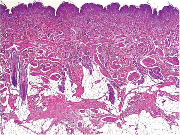
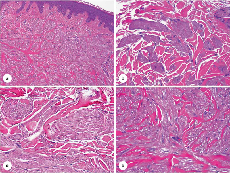
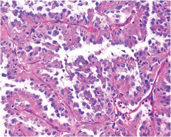
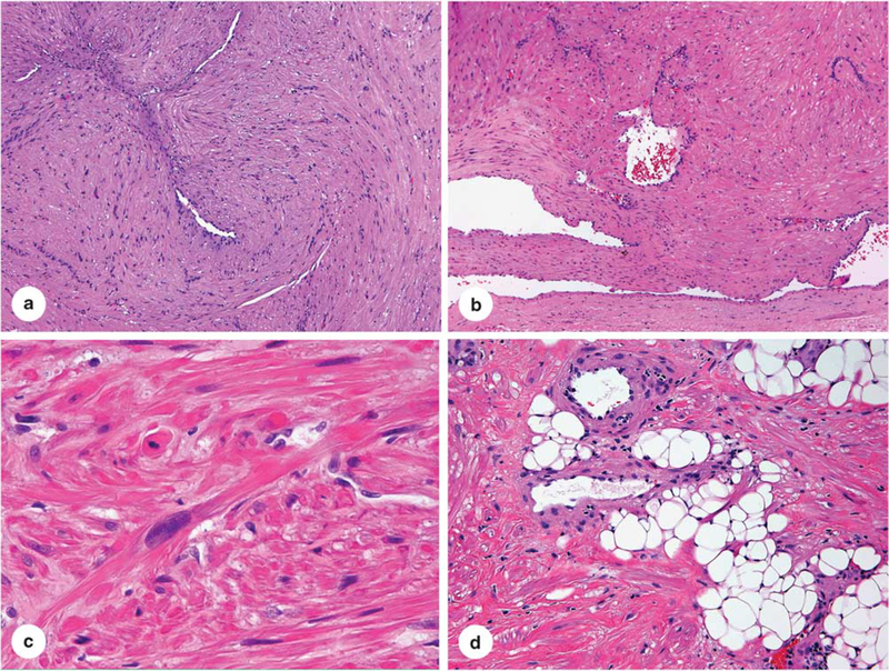
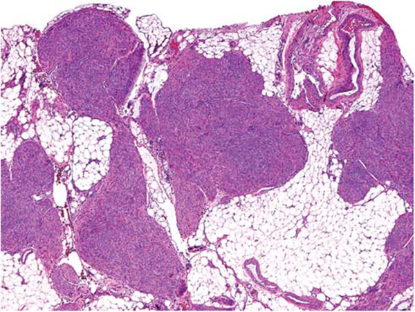
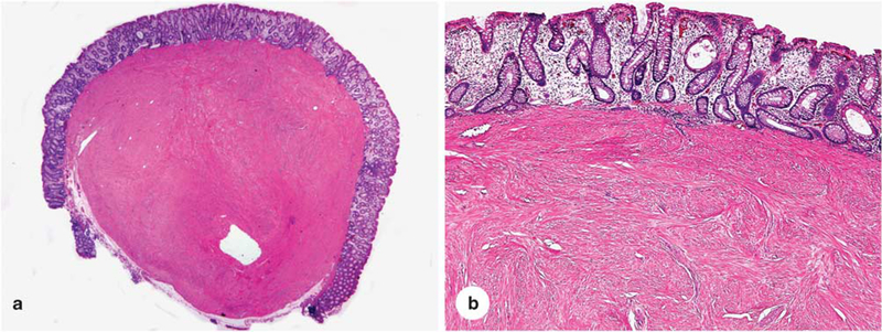
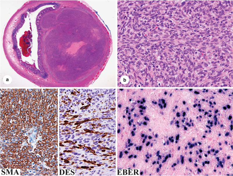
Smooth muscle tumors are here considered an essentially dichotomous group composed of benign leiomyomas and malignant leiomyosarcomas. Soft tissue smooth muscle tumors with both atypia and mitotic activity are generally diagnosed leiomyosarcomas acknowledging potential for metastasis. However, lesions exist that cannot be comfortably placed in either category, and in such cases the designation 'smooth muscle tumor of uncertain biologic potential' is appropriate. The use of this category is often necessary with limited sampling, such as needle core biopsies. Benign smooth muscle tumors include smooth muscle hamartoma and angioleiomyoma. A specific category of leiomyomas are estrogen-receptor positive ones in women. These are similar to uterine leiomyomas and can occur anywhere in the abdomen and abdominal wall. Leiomyosarcomas can occur at any site, although are more frequent in the retroperitoneum and proximal extremities. They are recognized by likeness to smooth muscle cells but can undergo pleomorphic evolution ('dedifferentiation'). Presence of smooth muscle actin is nearly uniform and desmin-positivity usual. This and the lack of KIT expression separate leiomyosarcoma from GIST, an important problem in abdominal soft tissues. EBV-associated smooth muscle tumors are a specific subcategory occurring in AIDS or post-transplant patients. These tumors can have incomplete smooth muscle differentiation but show nuclear EBER as a diagnostic feature. In contrast to many other soft tissue tumors, genetics of smooth muscle tumors are poorly understood and such diagnostic testing is not yet generally applicable in this histogenetic group. Leiomyosarcomas are known to be genetically complex, often showing 'chaotic' karyotypes including aneuploidy or polyploidy, and no recurrent tumor-specific translocations have been detected.
Conflict of interest statement
Disclosure/conflict of interest
The author declares no conflict of interest.
Figures


References
-
- Urbanek RW, Johnson WC. Smooth muscle hamartomas associated with Becker’s nevus. Arch Dermatol 1978;114:104–106. - PubMed
-
- Berger TG, Levin MW. Congenital smooth muscle hamartoma. J Am Acad Dermatol 1984;11:709–712. - PubMed
-
- Slifman NR, Harrist TJ, Rhodes AR. Congenital arrector pili hamartoma. A case report and review of the spectrum of Becker’s melanosis and pilar smooth-muscle hamartoma. Arch Dermatol 1985;121: 1034–1037. - PubMed
-
- Johnson MD, Jacobs AH. Congenital smooth muscle hamartoma. A report of six cases and review of the literature. Arch Dermatol 1989;125:820–822. - PubMed
-
- Zvulunov A, Rotem A, Merlob P, et al. Congenital smooth muscle hamartoma. Prevalence, clinical findings, and follow-up in 15 patients. Am J Dis Child 1990;144:782–784. - PubMed
Publication types
MeSH terms
Substances
Grants and funding
LinkOut - more resources
Full Text Sources
Other Literature Sources
