. 2013 Nov 25;203(4):575-83.
doi: 10.1083/jcb.201306012.

Atrophin-Rpd3 complex represses Hedgehog signaling by acting as a corepressor of CiR

Affiliations

Atrophin-Rpd3 complex represses Hedgehog signaling by acting as a corepressor of CiR

Zhao Zhang et al. J Cell Biol. .

Abstract

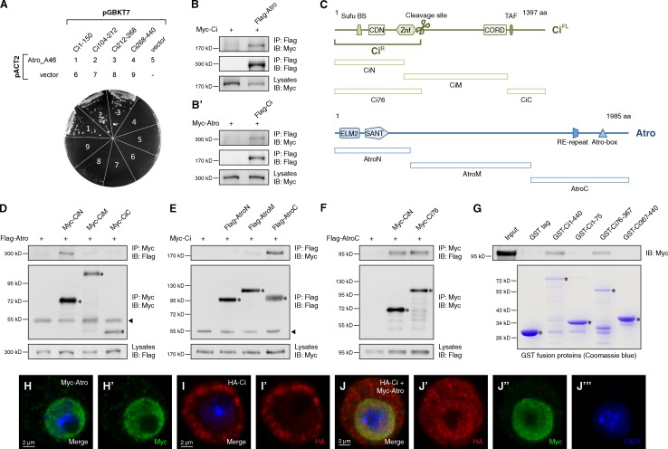
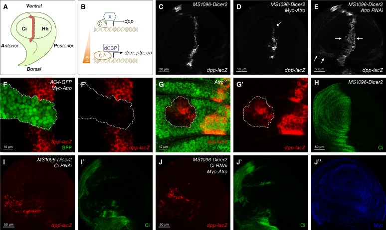
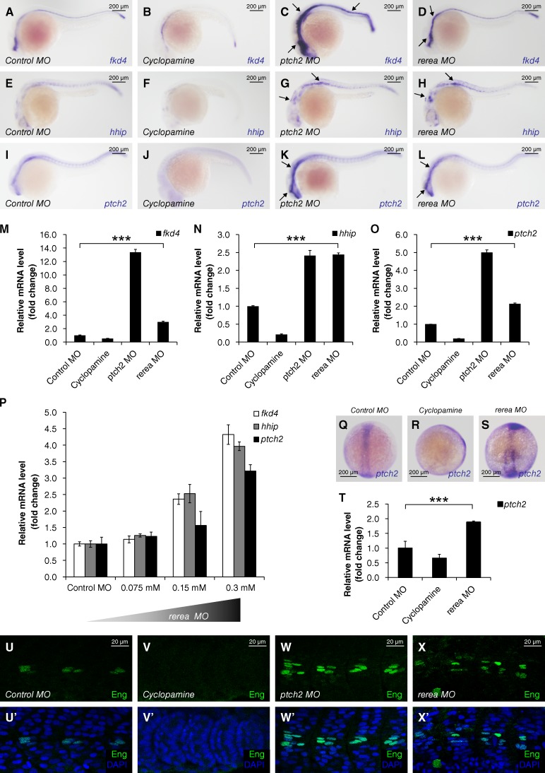
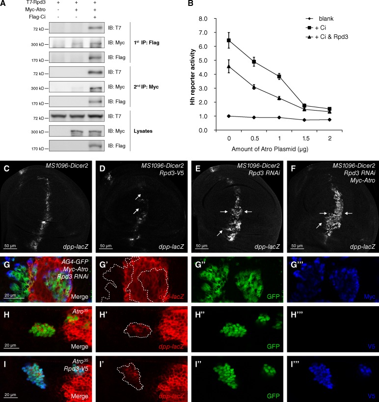
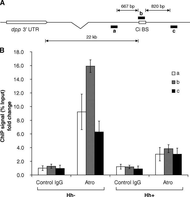
The evolutionarily conserved Hedgehog (Hh) signaling pathway is transduced by the Cubitus interruptus (Ci)/Gli family of transcription factors that exist in two distinct repressor (Ci(R)/Gli(R)) and activator (Ci(A)/Gli(A)) forms. Aberrant activation of Hh signaling is associated with various human cancers, but the mechanism through which Ci(R)/Gli(R) properly represses target gene expression is poorly understood. Here, we used Drosophila melanogaster and zebrafish models to define a repressor function of Atrophin (Atro) in Hh signaling. Atro directly bound to Ci through its C terminus. The N terminus of Atro interacted with a histone deacetylase, Rpd3, to recruit it to a Ci-binding site at the decapentaplegic (dpp) locus and reduce dpp transcription through histone acetylation regulation. The repressor function of Atro in Hh signaling was dependent on Ci. Furthermore, Rerea, a homologue of Atro in zebrafish, repressed the expression of Hh-responsive genes. We propose that the Atro-Rpd3 complex plays a conserved role to function as a Ci(R) corepressor.


Figures

Figure 1.
Figure 1.
AtroC binds to the CiN. (A) Yeast two-hybrid assay between Atro_A46 fragment and indicated Ci fragments. (B and B′) Western blots of immunoprecipitates (top two panels) or lysates (bottom) from S2 cells expressing the indicated proteins. (C) Schematic representation of domains and motifs in Atro and Ci proteins and their fragments used in subsequent coIP assay. (D–F) Western blots of immunoprecipitates (top two panels) or lysates (bottom) from S2 cells expressing the indicated proteins. (G) GST pull-down between Myc-tagged AtroC and GST or GST-tagged Ci fragments. (H–J‴) S2 cells expressing the indicated proteins were immunostained with HA (red), Myc (green) antibodies, and DAPI (blue) to visualize nuclei. In all blots, asterisks indicate the target proteins and arrowheads indicate IgG.
Figure 2.
Figure 2.
Atro is a repressor of Hh signaling pathway in D. melanogaster. (A) A cartoon of the wing discs from third-instar larva. The dashed line indicates the A/P compartment boundary. dpp expression region is shown in red. (B) Schematic shows that Hh gradient induces different target gene expression through transcriptional factor Ci. (C–E) A wild-type wing disc (C) or wing discs expressing Myc-tagged Atro (Myc-Atro; D) or Atro RNAi (E) with MS1096-Dicer2 Gal4 were immunostained to show the expression of dpp-lacZ. dpp-lacZ down-regulated in D (arrow) and up-regulated in E (arrows). (F and F′) A wing disc–expressing Myc-Atro with AG4-GFP was immunostained to show the expression of dpp-lacZ (red) and GFP (green). Atro-expressing clones (dashed lines) were marked by GFP-positive cells. (G and G′) A wing disc carrying Atro35 mutant clones (marked by GFP-positive cells in dashed lines) was immunostained to show the expression of dpp-lacZ (red) and GFP (green). (H) A MS1096-Dicer2 Gal4 wing disc was immunostained to show wild-type Ci staining. (I–J″) Wing discs expressing Ci RNAi alone or Ci RNAi and Myc-Atro together with MS1096-Dicer2 Gal4 were immunostained to show the expression of dpp-lacZ (red), Ci (green), and Myc-tagged Atro (blue). All of the phenotypes shown are stable and representative under each of the indicated genetic conditions.
Figure 3.
Figure 3.
The repressor function of Atro in Hh signaling is evolutionally conserved in zebrafish. (A–L) Expression of Hh-responsive genes in zebrafish embryos that were injected with the indicated MOs or treated with cyclopamine (10 µM) at 24 hpf. Up-regulated in situ stainings are marked by arrows. (M–O) Relative mRNA levels of fkd4, hhip, and ptch2 from 24 hpf zebrafish embryos indicated in A–L were revealed by real-time PCR (mean ± SD; n ≥ 3). (P) Relative mRNA levels of fkd4, hhip, and ptch2 from 24 hpf zebrafish embryos, which were injected with gradient concentrations of rerea MO. (Q–S) Expression of ptch2 in zebrafish embryos injected with the indicated MOs or treated with cyclopamine (100 µM) at bud stage (10 hpf). (T) Relative mRNA levels of ptch2 from bud stage embryos indicated in Q–S were revealed by real-time PCR (mean ± SD; n ≥ 3). (U–X’) Zebrafish embryos injected with the indicated MOs or treated with cyclopamine (10 µM) at 24 hpf were immunostained with Eng antibody (green) and DAPI (blue) to visualize nuclei. P-values in this figure were obtained by student’s t test between two groups (***, P < 0.001).
Figure 4.
Figure 4.
Atro recruits Rpd3 to repress Hh signaling. (A) S2 cells expressing the indicated proteins were harvested for the two-step immunoprecipitation and analyzed by Western blotting. (B) Rpd3 cooperates with Atro to repress Hh signaling in reporter assay in S2 cells (mean ± SD; n = 3). The whole DNA amount transfected in each group was normalized equal with blank vectors. (C–F) A wild-type wing disc (C) or wing discs expressing Rpd3-V5 (D), Rpd3 RNAi (E), or Rpd3 RNAi plus Myc-Atro (F) with MS1096-Dicer2 Gal4 were immunostained to show the expression of dpp-lacZ. The level of dpp-lacZ was reduced in Rpd3 overexpressing disc (D, arrows) and increased in Rpd3 knockdown disc (E, arrows). Atro overexpression failed to block the up-regulated expression of dpp-lacZ by Rpd3 knockdown (F, arrows). (G–G‴) A wing disc expressing Myc-Atro plus Rpd3 RNAi with AG4-GFP was immunostained to show the expression of dpp-lacZ– (red), GFP- (green), and Myc-tagged Atro (blue). (H–I‴) Wing discs carrying Atro35 clones or Atro35 plus Rpd3 overexpression were immunostained to show the expression of dpp-lacZ (red) and GFP (green). From G to I‴, clones were marked by GFP-positive cells (dashed lines).
Figure 5.
Figure 5.
Atro–Rpd3 complex associates with a Ci-binding site at the dpp locus in the absence of Hh signaling. (A) Schematic diagram of dpp locus and regions amplified with corresponding PCR primers to detect ChIP products. (B) ChIP for Atro around Ci binding site in dpp locus in Cl.8 cells with/without Hh treatment. Data of ChIP signals were normalized to 1/10 of input and showed as the fold change to the first group (mean ± SD; n = 3).

References

    1. Akimaru H., Chen Y., Dai P., Hou D.X., Nonaka M., Smolik S.M., Armstrong S., Goodman R.H., Ishii S. 1997. Drosophila CBP is a co-activator of cubitus interruptus in hedgehog signalling. Nature. 386:735–738 10.1038/386735a0 - DOI - PubMed
    1. Asai Y., Chan D.K., Starr C.J., Kappler J.A., Kollmar R., Hudspeth A.J. 2006. Mutation of the atrophin2 gene in the zebrafish disrupts signaling by fibroblast growth factor during development of the inner ear. Proc. Natl. Acad. Sci. USA. 103:9069–9074 10.1073/pnas.0603453103 - DOI - PMC - PubMed
    1. Aza-Blanc P., Ramírez-Weber F.A., Laget M.P., Schwartz C., Kornberg T.B. 1997. Proteolysis that is inhibited by hedgehog targets Cubitus interruptus protein to the nucleus and converts it to a repressor. Cell. 89:1043–1053 10.1016/S0092-8674(00)80292-5 - DOI - PubMed
    1. Charroux B., Freeman M., Kerridge S., Baonza A. 2006. Atrophin contributes to the negative regulation of epidermal growth factor receptor signaling in Drosophila. Dev. Biol. 291:278–290 10.1016/j.ydbio.2005.12.012 - DOI - PubMed
    1. Chen J.K., Taipale J., Cooper M.K., Beachy P.A. 2002. Inhibition of Hedgehog signaling by direct binding of cyclopamine to Smoothened. Genes Dev. 16:2743–2748 10.1101/gad.1025302 - DOI - PMC - PubMed

Publication types

MeSH terms