Comparative Study
. 2013 Sep:111:119-32.

Postulating a role for connective tissue elements in inferior oblique muscle overaction (an American Ophthalmological Society thesis)

Affiliations
Comparative Study

Postulating a role for connective tissue elements in inferior oblique muscle overaction (an American Ophthalmological Society thesis)

David Stager Jr et al. Trans Am Ophthalmol Soc. 2013 Sep.

Abstract

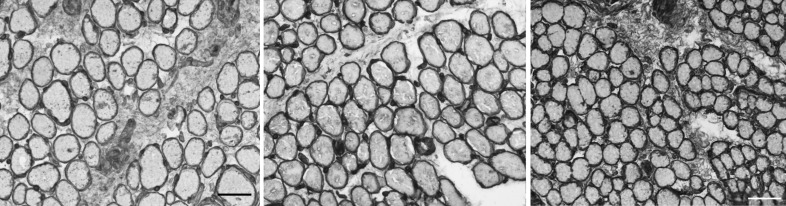
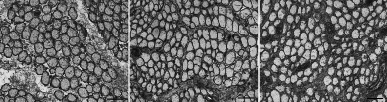
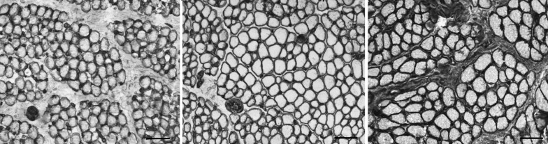
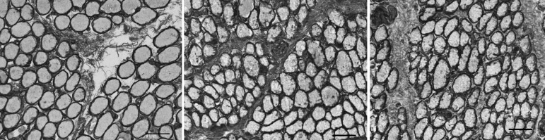
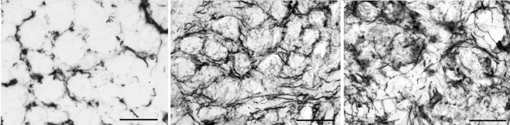
Purpose: To compare the localization and density of collagens I, IV, VI, and elastin, the major protein components of connective tissue, in the inferior oblique muscle of patients with overelevation in adduction and in controls and to characterize changes that develop following surgery. Biomechanical studies suggest that the connective tissue matrix plays a critical role in extraocular muscle function, determining tensile strength and force transmission during contraction.

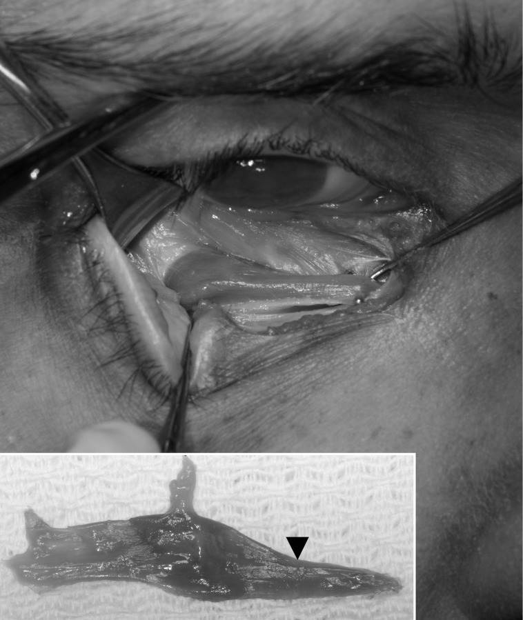
Methods: Prospective laboratory-based case-control study of inferior oblique muscle specimens from 31 subjects: 16 with primary inferior oblique overaction, 6 with craniofacial dysostosis, and 9 normal controls. Collagen I, IV, VI, and elastin were localized and quantified using immunohistochemical staining. Densities were compared using analysis of variance and post hoc comparisons.

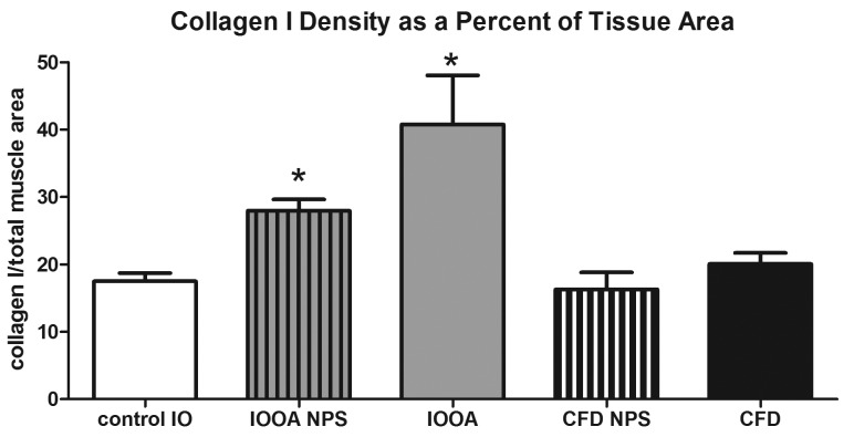
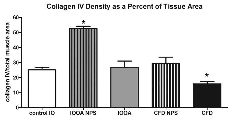
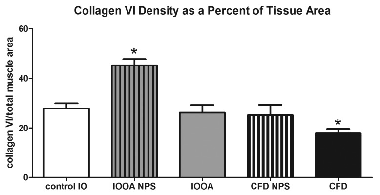
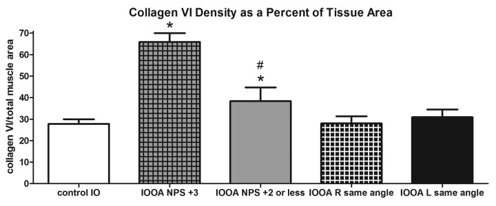
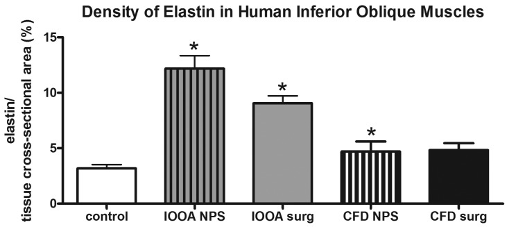
Results: In primary inferior oblique overaction, all connective tissue components in unoperated specimens were elevated compared to controls (P<.0001). Previously operated muscles showed normal levels of collagens IV and VI (P>.27) but increased collagen I. In unoperated craniofacial dysostosis specimens, only elastin was elevated (P=.03), whereas density of collagens IV and VI was lower in previously operated vs unoperated specimens (P=.015).

Conclusions: Elevated collagen and elastin levels in the cohort with primary inferior oblique overaction are consistent with the clinical finding of muscle stiffness. Contrarily, normal connective tissue densities in craniofacial dysostosis support the hypothesis that overelevation in this group reflects anomalous muscle vectors rather than tissue changes. Surgical intervention was associated with changes in the connective tissue matrix in both cohorts. These results have ramifications for treating patients with overelevation in adduction.


Figures

FIGURE 1
FIGURE 1
Gross anatomy of the inferior oblique muscle, which is isolated with a muscle hook. The inset shows characteristic narrowing of the nasal portion of the muscle.
FIGURE 2
FIGURE 2
Immunohistochemical visualization of collagen in normal inferior oblique muscles from children. Collagen I in control subject 5 (left); collagen IV in control subject 3 (center); and collagen VI in control subject 3 (right) (stained for collagen I, IV, and VI; bar = 50 μm).
FIGURE 3
FIGURE 3
Expression of collagen I in inferior oblique muscles. Left, normal (control subject 3). Center, a patient who had no prior strabismus surgery (primary inferior oblique overaction subject 10). Right, a patient who had prior strabismus surgery (primary inferior oblique overaction subject 7) (stained for collagen I; bar = 50 μm).
FIGURE 4
FIGURE 4
Mean density of collagen I in inferior oblique muscles. Data is expressed as percent expression of collagen I per muscle area and presented as the mean ±1 standard error of the mean (SEM). Asterisk (*) indicates significantly different from control values at P<.05. CFD, craniofacial dysostosis; IO, inferior oblique; IOOA, inferior oblique overaction; NPS, no prior surgery.
FIGURE 5
FIGURE 5
Expression of collagen IV in inferior oblique muscles. Left, normal (control subject 1). Center, a patient who had no prior strabismus surgery (primary inferior oblique overaction subject 10). Right, a patient who had prior strabismus surgery (primary inferior oblique overaction subject 8) (stained for collagen IV; bar = 50 μm).
FIGURE 6
FIGURE 6
Mean density of collagen IV in inferior oblique muscles. Data is expressed as percent expression of collagen IV per muscle area and presented as the mean ±1 standard error of the mean (SEM). Asterisk (*) indicates significantly different from control values at P<.05. CFD, craniofacial dysostosis; IO, inferior oblique; IOOA, inferior oblique overaction; NPS, no prior surgery.
FIGURE 7
FIGURE 7
Expression of collagen VI in inferior oblique muscles. Left, normal (control subject 5). Center, a patient who had no prior strabismus surgery (primary inferior oblique overaction subject 11). Right, a patient who had prior strabismus surgery (primary inferior oblique overaction subject 5). (stained for collagen VI; bar = 50 μm).
FIGURE 8
FIGURE 8
Mean density of collagen VI in inferior oblique muscles. Data is expressed as percent expression of collagen VI per muscle area and presented as the mean ±1 standard error of the mean (SEM). Asterisk (*) indicates significantly different from control values at P<.05. CFD, craniofacial dysostosis; IO, inferior oblique; IOOA, inferior oblique overaction; NPS, no prior surgery.
FIGURE 9
FIGURE 9
Mean density of collagen VI in inferior oblique muscles. Results are compared from controls, patients whose angle of misalignment varied in their two eyes, and those where similar angles of misalignment were seen. Data is expressed as percent expression of collagen VI per muscle area and presented as the mean ±1 standard error of the mean (SEM). Asterisk (*) indicates significantly different from control values at P<.05; # sign indicates significantly different from contralateral side at P<.05. IO, inferior oblique; IOOA, inferior oblique overaction; NPS, no prior surgery.
FIGURE 10
FIGURE 10
Expression of elastin in inferior oblique muscles. Left, normal (control subject 5). Center, a patient who had no prior strabismus surgery (primary inferior oblique overaction subject 12). Right, a patient who had prior strabismus surgery (primary inferior oblique overaction subject 3) (stained for elastin; bar = 20 μm).
FIGURE 11
FIGURE 11
Mean density of elastin in inferior oblique muscles. Data is expressed as percent expression of collagen VI per muscle area and presented as the mean ±1 standard error of the mean (SEM). Asterisk (*) indicates significantly different from control values at P<.05. CFD, craniofacial dysostosis; IO, inferior oblique; IOOA, inferior oblique overaction; NPS, no prior surgery.

References

    1. Kushner BJ. Multiple mechanisms of extraocular muscle “overaction”. Arch Ophthalmol. 2006;124(5):680–688. - PubMed
    1. Kushner BJ. Incomitant strabismus: Does extraocular muscle form denote function? Arch Ophthalmol. 2010;128(12):1604–1609. - PubMed
    1. Von Noorden GK. Atlas of Strabismus. 4th ed. StLouis: CV Mosby Co; 1983.
    1. Hertle RW, National Eye Institute Sponsored Classification of Eye Movement Abnormalities and Strabismus Working Group A next step in naming and classification of eye movement disorders and strabismus. J AAPOS. 2002;6(4):201–202. - PubMed
    1. American Academy of Ophthalmology . Pediatric Ophthalmology and Strabismus. San Francisco: American Academy of Ophthalmology; 2006.

Publication types

MeSH terms