. 2013 Dec 30;8(12):e84273.
doi: 10.1371/journal.pone.0084273. eCollection 2013.

Characterization of antigen-specific B cells using nominal antigen-coated flow-beads

Affiliations

Characterization of antigen-specific B cells using nominal antigen-coated flow-beads

Nicolas Degauque et al. PLoS One. .

Erratum in

  • PLoS One. 2014;9(7):e102845. Ngono, Annie Elong [corrected to Elong Ngono, Annie]

Abstract

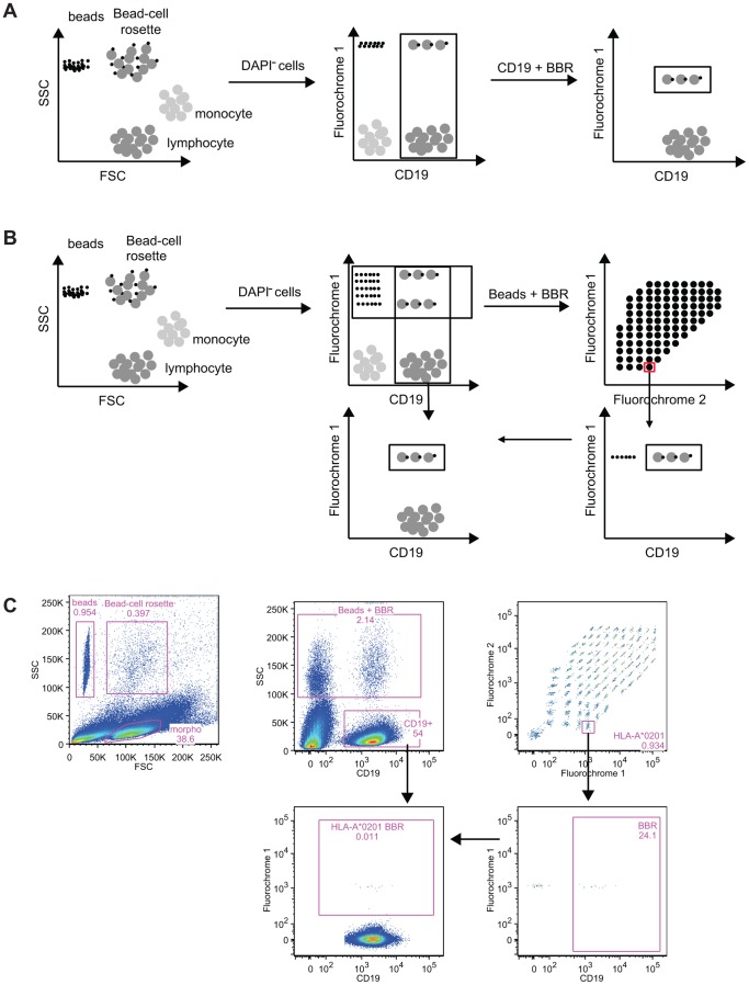
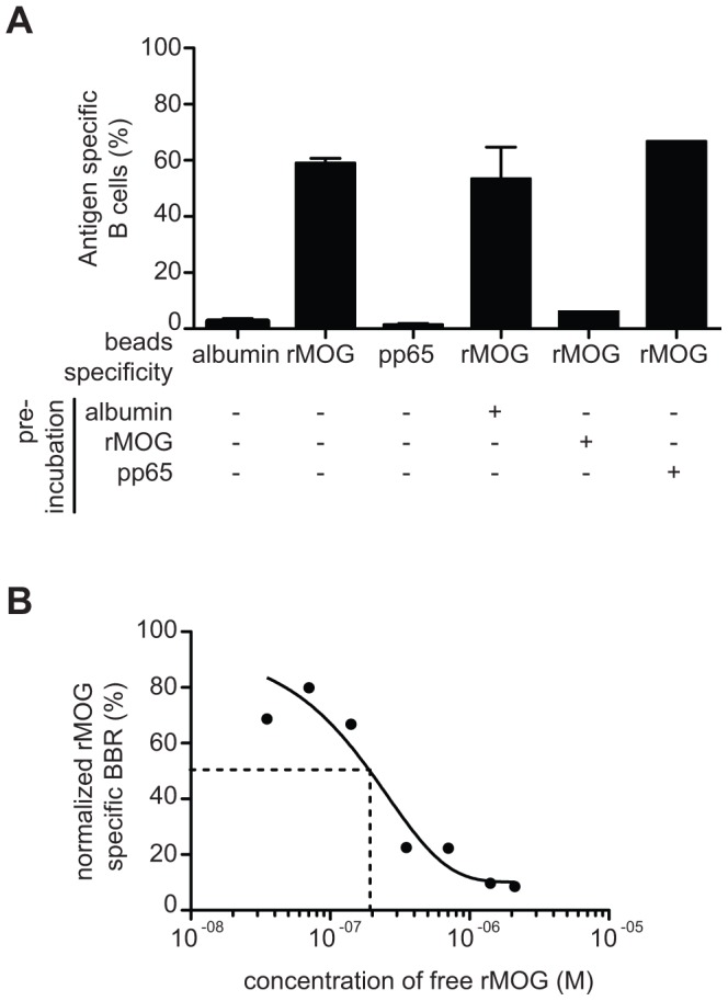
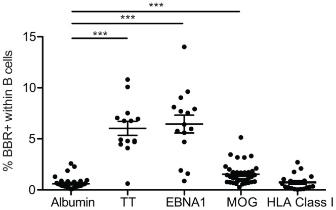
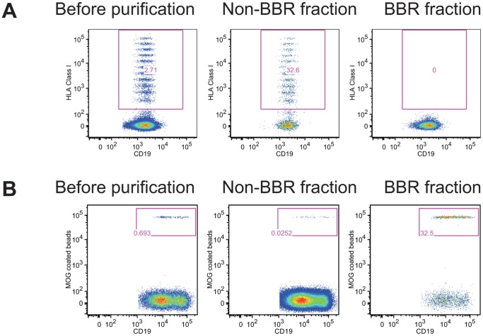
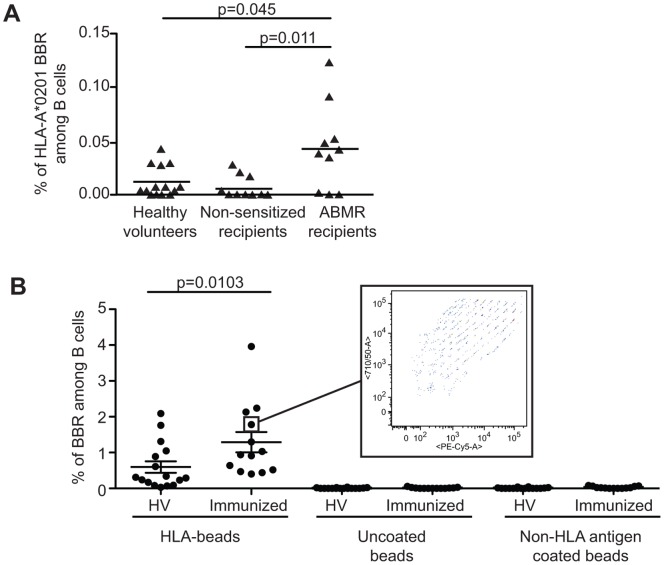
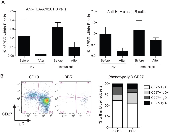
In order to characterize the reactivity of B cells against nominal antigens, a method based on the coupling of antigens onto the surface of fluorescent core polystyrene beads was developed. We first demonstrate that murine B cells with a human MOG-specific BCR are able to interact with MOG-coated beads and do not recognize beads coated with human albumin or pp65. B cells purified from human healthy volunteer blood or immunized individuals were tested for their ability to interact with various nominal antigens, including viral, vaccine, self and alloantigens, chosen for their usefulness in studying a variety of pathological processes. A substantial amount of B cells binding self-antigen MOG-coated beads can be detected in normal blood. Furthermore, greater frequencies of B cell against anti-Tetanic Toxin or anti-EBNA1 were observed in primed individuals. This method can reveal increased frequencies of anti-HLA committed B cells in patients with circulating anti-HLA antibodies compared to unsensitized patients and normal individuals. Of interest, those specific CD19 cells were preferentially identified within CD27(-)IgD(+) (i-e naïve) subset. These observations suggest that a broad range of medical situations could benefit from a tool that allows the detection, the quantification and the characterization of antigen-specific blood B cells.


Conflict of interest statement

Competing Interests: The authors have declared that no competing interests exist.

Figures

Figure 1
Figure 1. Principle of the method of identification of antigen-specific B cells.
After co-incubation, lymphocytes, antigen covered beads and the beads’ B cell rosettes are gated based on their forward scatter and side scatter. After exclusion of the DAPI+ cells, B cells and beads-B cell rosettes are identified based on CD19 expression and the beads’ internal fluorochrome. Specificity of B cell recognition is determined by gating on beads and beads’ B cell rosettes (A) or after the identification of the nominal antigen through the use of the unique ratio of the two internal fluorochromes (B). In the latter, for each nominal antigen, a gate that encompassed beads and B cell rosettes is created followed by the identification of the B cells. Frequency of B cells bound to HLA class I of interest is finally evaluated. Bead-based method allows the detection of antigen-specific B cells. (C). An example of the identification of beads, Bead-cell rosette and lymphocyte is shown. After exclusion of dead cells, the use of the marker CD19 allows the identification of B lymphocyte and a mix of beads and BBR. Thanks to the ratio of two fluorochromes, antigen coated on the beads can be then identified. Beads are excluded using the expression of CD19. A Boolean gate is used to assess the frequency of B cells specific of a given antigen within the whole B cell population.
Figure 2
Figure 2. Bead-based method allows the detection of antigen-specific B cells.
(A) B cells purified from Tg mice were incubated with human albumin, MOG1–125 or pp65 coated beads and the frequency of antigen specific B cells was quantified. The B cells were preincubated with soluble human Albumin, MOG1–125 or pp65 before incubation with MOG1–125 coated beads. Data are presented as mean ± sem B). B cells purified from Tg mice were preincubated with increasing doses of soluble MOG1–125 before incubation with MOG1–125 coated beads. The experiments were repeated 3 times and similar results were obtained.
Figure 3
Figure 3. B cells from healthy volunteers exhibit a broad range of reactivity.
Purified B cells from healthy volunteers were tested for their reactivity to albumin (n = 38), Tetanus Toxin (n = 14), EBNA1 (n = 15), MOG1–125 (n = 38) and a panel of 97 HLA class I molecules (n = 19). ***p<0.001 (Kruskall-Wallis follow by a Dunn’s post hoc test using albumin settings as reference group).
Figure 4
Figure 4. Separation of B cells interacting with nominal antigen and unbound B cells.
Purified B cells were incubated with single HLA class I coated beads (A) or MOG1–125 coated beads (B) before being subjected to cell separation using an ARIA FACS-sorter (A) or magnet based purification (B). Frequency of B cells interacting with nominal antigens is shown before purification and in the positive and in the negative fraction. One representative out of three experiments with cells from different donors is shown.
Figure 5
Figure 5. Enhanced frequency of anti-HLA B cell in immunized patients.
A. Using single HLA-A*0201 coated beads, the frequency of B cells specific to HLA-A*0201 allele was assessed in the blood of sensitized transplant recipients with histologically proven antibody mediated rejection (ABMR; n = 10), non-sensitized stable transplant recipients (n = 9) and healthy volunteers (n = 14). Sensitized patients exhibit a significant increase in the frequency of HLA-A*0201 specific B cells compared to non-sensitized patients and healthy volunteers. p value are mentioned (Kruskall-Wallis follow by a Dunn’s post hoc test) B. B cells bound to single HLA class I coated beads (HLA-beads), to negative control (NC) and positive control (PC) were analyzed in HV (n = 16) and Immunized kidney recipients (n = 13). NC and PC beads were included by the manufacture in the single HLA class I kit. According to the manufacture, NC beads are beads saturated with ovalbumin and PC beads are coated with human IgG1. A broad range of single HLA class I were recognized as shown in the insert, a pattern observed for B cells from all tested patients. p value is indicated (Mann-Whitney test).
Figure 6
Figure 6. The interaction between CD19+ cells and HLA class I coated beads is not restricted by the HLA class I allele only and BBR are not restricted to the memory compartment.
A. CD19+ cells were pre-incubated with a cocktail of HLA-A*0201 multimer (HLA-A*0201/MP58–66, HLA-A*0201/HCw1, HLA-A*0201/pp65) followed by the incubation with HLA Class I coated beads. Frequency of CD19+ cells specific of HLA-A*0201 coated beads and of HLA class I coated beads were analyzed before and after coincubation with the cocktail of HLA-A*0201 multimer for healthy volunteers (n = 6) and immunized patients (n = 3). Data are presented as mean ± sem. B. B cells were stained with anti-CD19, anti-CD27 and anti-IgD antibodies prior to incubation with single HLA coated beads. Phenotype of B cells and BBR were analyzed based on the expression of CD27 and IgD. 4 populations were identified (CD27IgD+, naïve B cells; CD27+IgD+, non-switched memory B cells; CD27+IgD, switched memory B cells; CD27IgD); CD27IgD (late memory B cells). Representative phenotype of B cells and single HLA class I coated beads is shown as well as a summary of 5 immunized patients (mean).

References

    1. Hawker K, O’Connor P, Freedman MS, Calabresi PA, Antel J, et al. (2009) Rituximab in patients with primary progressive multiple sclerosis: Results of a randomized double-blind placebo-controlled multicenter trial. Ann Neurol 66: 460–471 10.1002/ana.21867 - DOI - PubMed
    1. Brulhart L, Ciurea A, Finckh A, Notter A, Waldburger JM, et al. (2006) Efficacy of B cell depletion in patients with rheumatoid arthritis refractory to anti-tumour necrosis factor agents: an open-label observational study. Annals of the Rheumatic Diseases 65: 1255–1257 10.1136/ard.2005.051169 - DOI - PMC - PubMed
    1. Iwata Y, Matsushita T, Horikawa M, DiLillo DJ, Yanaba K, et al. (2011) Characterization of a rare IL-10-competent B-cell subset in humans that parallels mouse regulatory B10 cells. Blood 117: 530–541 10.1182/blood-2010-07-294249 - DOI - PMC - PubMed
    1. Yanaba K, Bouaziz JD, Matsushita T, Tsubata T, Tedder TF (2009) The Development and Function of Regulatory B Cells Expressing IL-10 (B10 Cells) Requires Antigen Receptor Diversity and TLR Signals. The Journal of Immunology 182: 7459–7472 10.4049/jimmunol.0900270 - DOI - PMC - PubMed
    1. Frommer F, Heinen TJAJ, Wunderlich FT, Yogev N, Buch T, et al. (2008) Tolerance without clonal expansion: self-antigen-expressing B cells program self-reactive T cells for future deletion. The Journal of Immunology 181: 5748–5759. - PubMed

Publication types

MeSH terms