. 2013 Dec 26;8(12):e84286.
doi: 10.1371/journal.pone.0084286. eCollection 2013.

Secretory activity is rapidly induced in stigmatic papillae by compatible pollen, but inhibited for self-incompatible pollen in the Brassicaceae

Affiliations

Secretory activity is rapidly induced in stigmatic papillae by compatible pollen, but inhibited for self-incompatible pollen in the Brassicaceae

Darya Safavian et al. PLoS One. .

Abstract

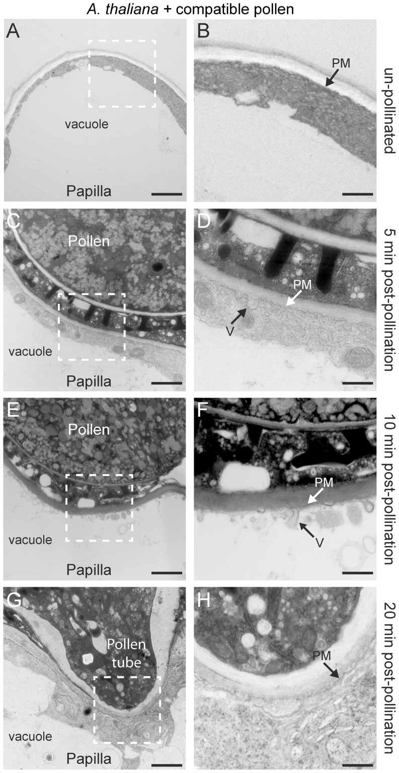
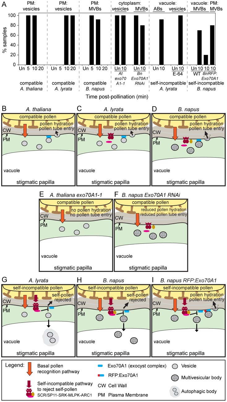
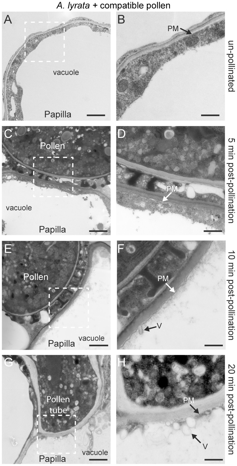
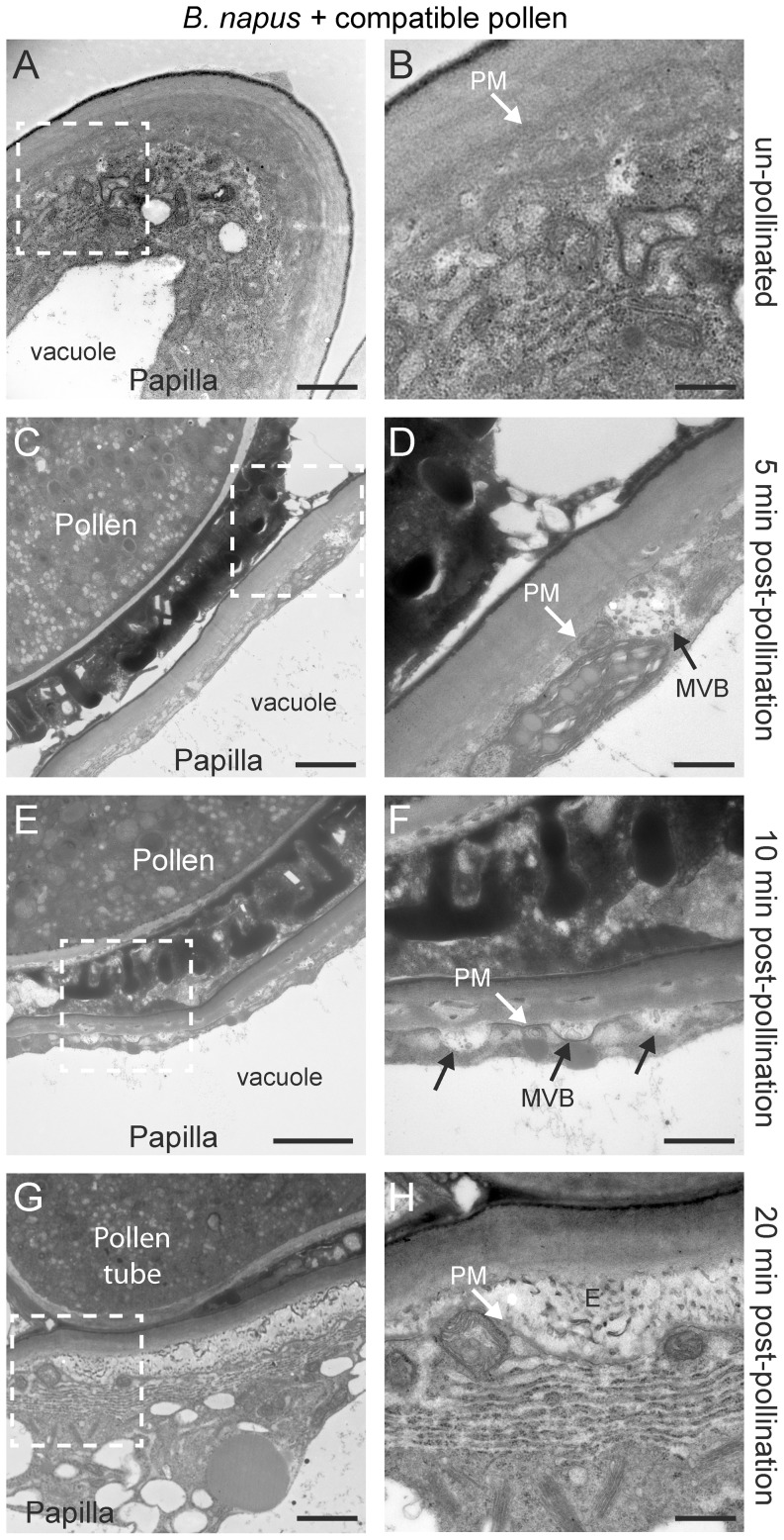
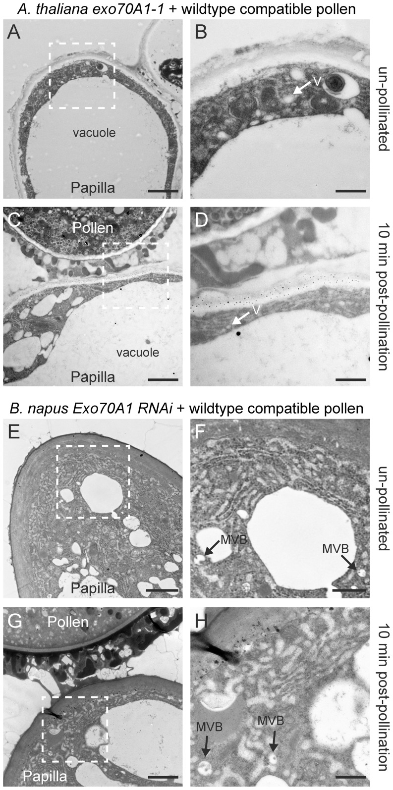
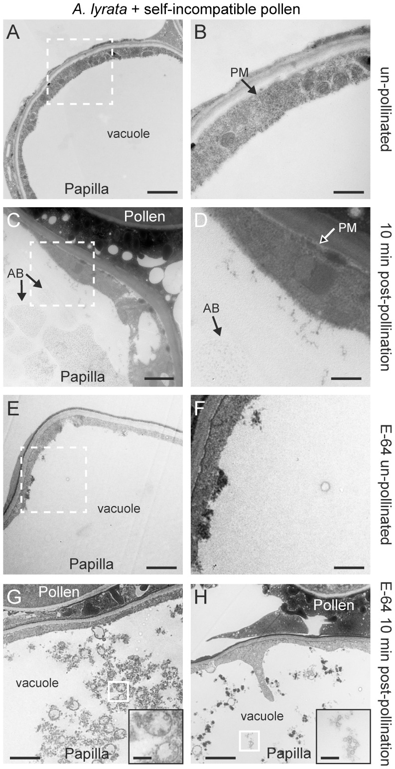
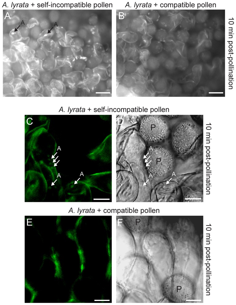
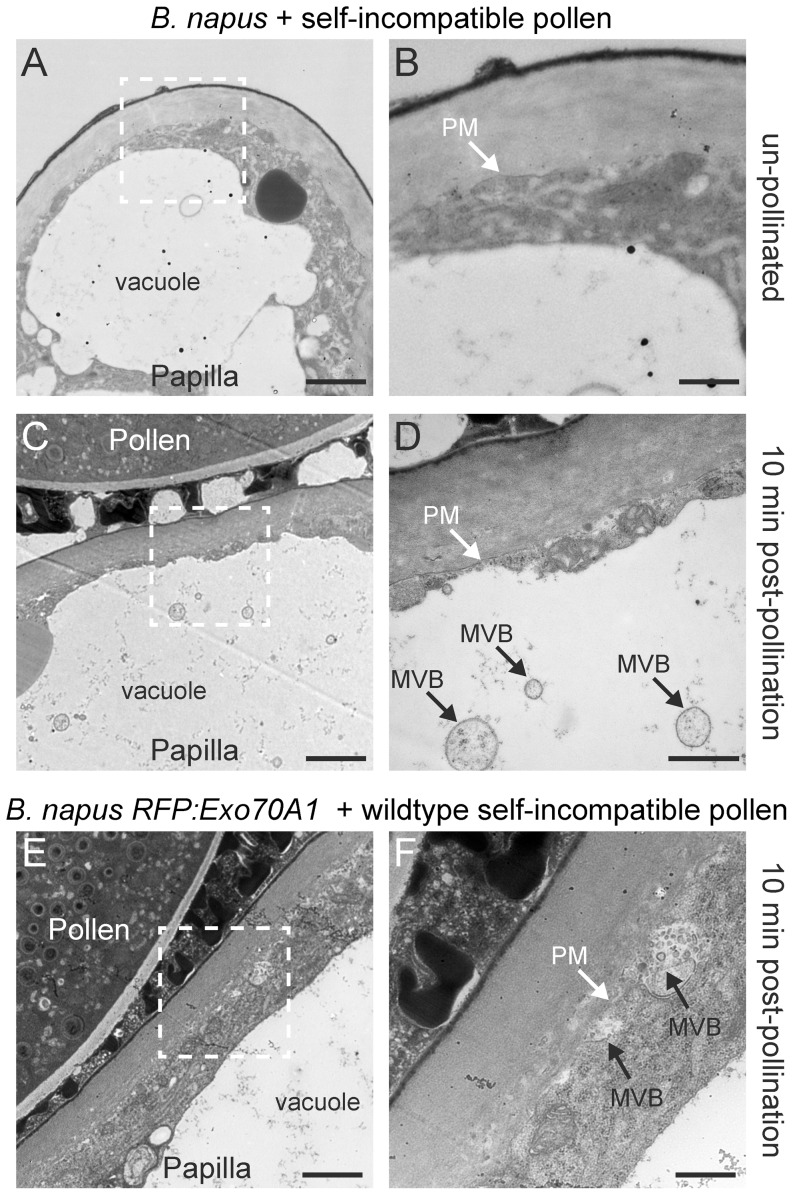
[In the Brassicaceae, targeted exocytosis to the stigmatic papillar plasma membrane under the compatible pollen grain is hypothesized to be essential for pollen hydration and pollen tube penetration. In contrast, polarized secretion is proposed to be inhibited in the stigmatic papillae during the rejection of self-incompatible pollen. Using transmission electron microscopy (TEM), we performed a detailed time-course of post-pollination events to view the cytological responses of the stigmatic papillae to compatible and self-incompatible pollinations. For compatible pollinations in Arabidopsis thaliana and Arabidopsis lyrata, vesicle secretion was observed at the stigmatic papillar plasma membrane under the pollen grain while Brassica napus stigmatic papillae appeared to use multivesicular bodies (MVBs) for secretion. Exo70A1, a component of the exocyst complex, has been previously implicated in the compatible pollen responses, and disruption of Exo70A1 in both A. thaliana and B. napus resulted in a loss of secretory vesicles/MVBs at the stigmatic papillar plasma membrane. Similarly, for self-incompatible pollinations, secretory vesicles/MVBs were absent from the stigmatic papillar plasma membrane in A. lyrata and B. napus; and furthermore, autophagy appeared to be induced to direct vesicles/MVBs to the vacuole for degradation. Thus, these findings support a model where the basal pollen recognition pathway in the stigmatic papilla promotes exocytosis to accept compatible pollen, and the basal pollen recognition pathway is overridden by the self-incompatibility pathway to prevent exocytosis and reject self-pollen.


Conflict of interest statement

Competing Interests: The authors have declared that no competing interests exist.

Figures

Figure 1
Figure 1. TEM images of A. thaliana Col-0 stigmatic papillae in response to self-compatible pollen.
(A, B) Unpollinated stigmatic papilla. Secretory activity was not observed at the papillar plasma membrane (PM) in 10/10 samples. (C, D) Stigmatic papilla at 5 min post-pollination. Vesicles (V) were observed to be fusing to the plasma membrane (PM) underneath the pollen contact site in 25/25 samples. (E, F) Stigmatic papilla at 10 min post-pollination. Vesicles (V) continue to fuse to the plasma membrane (PM) underneath the pollen contact site in 25/25 samples. (G, H) Pollen tube penetration into the stigmatic papilla at 20 min post-pollination. Vesicles were no longer observed at the stigmatic papillar plasma membrane (PM) in 25/25 samples. The white boxed areas in (A, C, E, G) are shown in the (B, D, F, H), respectively. Scale bars (A, C, E, G) 1.5 µm; (B, D, F, H) 500 nm
Figure 2
Figure 2. Summary of the stigmatic papillar responses to compatible and self-incompatible pollen.
(A) Percentage of samples with the main ultrastructural features observed in the TEM images. Corresponding TEM images are shown in Figures 1, 4–7 and 9. Abbreviations: Un  =  unpollinated; MVBs  =  multivesicular bodies; ABs  =  autophagic bodies; PM  =  plasma membrane. (B-D) Models for compatible pollen responses (based on this study and cited references). Under the compatible pollen contact site, an unknown basal pollen recognition pathway is activated in the stigmatic papilla. This leads to the assembly of the exocyst complex with Exo70A1 and plasma membrane docking of vesicles in Arabidopsis species (B, C) or MVBs in B. napus (D). Following vesicle/MVB fusion to the plasma membrane by SNAREs, unknown cargo are released to facilitate water release for pollen hydration and cell wall expansion for pollen tube entry (pollen is accepted). (E, F) Models for loss of Exo70A1 function in the stigmatic papilla (based on this study and cited references). Disrupting Exo70A1 expression prevents plasma membrane docking of vesicles in A. thaliana exo70A1-1 (E) or MVBs in B. napus Exo70A1 RNAi (F). Consequently, vesicles/MVBs accumulate in the cytoplasm; cargo needed for accepting the compatible pollen are not delivered to the plasma membrane; and this leads to pollen rejection. (G, H) Models for self-incompatible pollen responses (based on this study and cited references). With self-incompatible pollen, the self-incompatibility pathway is activated in the stigmatic papilla and overrides the basal pollen recognition pathway by inhibiting Exo70A1 and vesicle/MVB docking. The vesicles in A. lyrata (G) or MVBs in B. napus (H) are redirected to the vacuole via autophagy for degradation. Consequently, pollen hydration and pollen tube penetration are prevented (self-pollen is rejected). (I) Model for the partial breakdown of self-incompatibility through the expression of RFP:Exo70A1 in B. napus . MVBs are able to dock at the plasma membrane despite the activation of the self-incompatibility pathway, and this leads to pollen acceptance (pollen hydration and pollen tube entry occur . RFP:Exo70A1 may potentially cause this phenotype through increased Exo70A1 levels or by interference with the ARC1 interaction.
Figure 3
Figure 3. TEM images of clathrin-coated vesicles in the A. thaliana root tip cells.
(A-C) The root tips from 6-day-old A. thaliana seedlings were observed as a reference for clathrin-coated vesicles (CCV) in the plant endocytic pathway. Clathrin-coated vesicles were observed adjacent to the plasma membrane in the root tip cells in 25/25 samples. Scale bars (A-C) 100 nm.
Figure 4
Figure 4. TEM images of A. lyrata stigmatic papillae in response to cross-compatible pollen.
(A, B) Unpollinated stigmatic papilla. Secretory activity was not observed at the papillar plasma membrane (PM) in 10/10 samples. (C, D) Stigmatic papilla at 5 min post-pollination. Secretory activity was not observed at the papillar plasma membrane (PM) in 10/10 samples. (E, F) Stigmatic papilla at 10 min post-pollination. Vesicles (V) were observed to be fusing to the plasma membrane (PM) underneath the pollen contact site in 25/25 samples. (G-H) Pollen tube penetration into the stigmatic papilla at 20 min post-pollination. Vesicles (V) were observed at the papillar plasma membrane (PM) beneath the pollen tube tip in 25/25 samples. The white boxed areas in (A, C, E, G) are shown in the (B, D, F, H), respectively. Scale bars (A, C, E, G) 1.5 µm; (B, D, F, H) 500 nm.
Figure 5
Figure 5. TEM images of B. napus Westar stigmatic papillae in response to self-compatible pollen.
(A, B) Unpollinated stigmatic papilla. Secretory activity was not observed at the papillar plasma membrane (PM) in 10/10 samples. (C, D) Stigmatic papilla at 5 min post-pollination showing an MVB fusing to the plasma membrane (PM). MVBs at the plasma membrane were observed in 10/10 samples. (E, F) Stigmatic papilla at 10 min post-pollination showing several MVBs fusing to the plasma membrane (PM) underneath the pollen contact site. MVBs at the plasma membrane were observed in 23/25 samples. For 2/25 samples, vesicles were observed to be fusing to the plasma membrane (Figure S2A, B). (G, H) Pollen tube penetration into the stigmatic papilla at 20 min post-pollination. The material under the papillar cell wall appears to be the exosomes (E) released from the MVBs. This pattern was observed in 15/15 samples. The white boxed areas in (A, C, E, G) are shown in the (B, D, F, H), respectively. Scale bars (A, C, E, G) 1.5 µm; (B, D, F, H) 500 nm.
Figure 6
Figure 6. TEM images of A. thaliana exo70A1-1 and B. napus Westar Exo70A1 RNAi stigmatic papillae in response to wild-type compatible pollen.
(A, B) Unpollinated stigmatic papilla from the A. thaliana exo70A1-1 mutant. Some vesicle (V) accumulation was observed in the cytoplasm in 10/10 samples. (C, D) Stigmatic papilla from the A. thaliana exo70A1-1 mutant at 10 min following pollination with compatible A. thaliana Col-0 pollen. An accumulation of secretory vesicles (V) in the papillar cytoplasm was observed under the pollen contact site in 10/10 samples. (E, F) Unpollinated stigmatic papilla from the B. napus Westar Exo70A1 RNAi R2 line. Some accumulation of MVBs was observed in the cytoplasm in 10/10 samples. (G, H) Stigmatic papilla from the B. napus Westar Exo70A1 RNAi R2 line at 10 min following pollination with compatible B. napus Westar pollen. Some MVBs were observed in the papillar cytoplasm in 8/10 samples. For 2/10 samples, MVBs were observed in the cytoplasm and fusing to the plasma membrane (Figure S2C, D) which is consistent with these plants displaying an incomplete knockout phenotype . The white boxed areas in (A, C, E, G) are shown in the (B, D, F, H), respectively. Scale bars (A, C, E, G) 1.5 µm; (B, D, F, H) 500 nm.
Figure 7
Figure 7. TEM images of A. lyrata stigmatic papillae in response to self-incompatible pollen.
(A, B) Unpollinated stigmatic papilla. Secretory activity was not observed at the papillar plasma membrane (PM), and the vacuole was largely clear (i.e no autophagic bodies were visible) in 10/10 samples. (C, D) Stigmatic papilla at 10 min post-pollination with self-incompatible pollen. No secretory activity was observed at the papillar plasma membrane (PM). Structures that may represent autophagic bodies (AB) were observed in the vacuole in 23/25 samples. In 2/25 samples, these structures were not visible in the vacuole. (E, F) Unpollinated stigmatic papilla treated with the E-64 inhibitor. The vacuole was largely clear (i.e. no autophagic bodies or vesicles were visible) in 10/10 samples. (G, H) Stigmatic papillae treated with the E-64 inhibitor, at 10 min post-pollination with self-incompatible pollen. Un-degraded vesicles were observed in the vacuole in 10/10 samples. The white boxed areas in (A, C, E) are shown in the (B, D, F), respectively. The white boxed areas in (G) and (H) are shown in the insets in the bottom right hand corners. Scale bars (A, C, E, G, H) 1.5 µm; (B, D, F) 500 nm, Insets for (G, H) 300 nm.
Figure 8
Figure 8. Autophagosomes in A. lyrata stigmatic papillae in response to self-incompatible pollen.
(A, B) Florescence microscopy images of MDC stained A. lyrata stigmatic papillae at 10 min post-pollination. Fluorescent signals that may represent autophagosomes were seen in the A. lyrata stigmatic papillae following a self-incompatible pollination (A) in 10/10 samples, but not observed after a cross-compatible pollination (B) in 10/10 samples. (C-F) Confocal microscopy images of transgenic A. lyrata GFP:ATG8a stigmatic papillae at 10 min post-pollination. GFP:ATG8a is a marker for autophagy induction, and GFP signals marking potential autophagosomes were observed in the stigmatic papillae following a self-incompatible pollination (C) in 10/10 samples (corresponding DIC image is shown in D). Punctate GFP signals were not detected within the stigmatic papillae following a cross-compatible pollination (E) in 10/10 samples (corresponding DIC image is shown in F). All samples, including wild-type untransformed A. lyrata stigmatic papillae showed background fluorescence from the cell wall. A  =  autophagosomes; P =  pollen. Scale bars (A, B) 50 µm; (C-F) 10 µm.
Figure 9
Figure 9. TEM images of B. napus W1 stigmatic papillae in response to self-incompatible pollen.
(A, B) Unpollinated B. napus W1 stigmatic papilla. Secretory activity was not observed at the papillar plasma membrane (PM), and the vacuole was largely clear (no MVBs were visible) in 10/10 samples. (C, D) B. napus W1 stigmatic papilla at 10 min post-pollination with self-incompatible pollen. Secretory activity was not observed at the papillar plasma membrane (PM). Instead, MVBs were observed in the vacuole in 7/10 samples. For 3/10 samples, MVBs were observed in the cytoplasm near the vacuole (Figure S2E, F). (E, F) Transgenic B. napus RFP:Exo70A1 W1 stigmatic papilla at 10 min post-pollination with self-incompatible pollen. These plants were previously found to have a partial breakdown of the self-incompatibility response due to RFP:Exo70A1 expression . Consistent with this partial phenotype, MVBs were observed to be fusing to the plasma membrane in 8/10 samples as shown in (F), and MVBs were also observed in the vacuole in 2/10 samples (Figure S2G, H). The white boxed areas in (A, C, E) are shown in the (B, D, F), respectively. Scale bars (A, C, E) 1.5 µm; (B, D, F) 500 nm.

Comment in

References

    1. Chapman LA, Goring DR (2010) Pollen-pistil interactions regulating successful fertilization in the Brassicaceae. J Exp Bot 61: 1987–1999. - PubMed
    1. Dickinson H (1995) Dry stigmas, water and self-incompatibility in Brassica . Sex Plant Reprod 8: 1–10.
    1. Hiscock SJ, Allen AM (2008) Diverse cell signalling pathways regulate pollen-stigma interactions: the search for consensus. New Phytol 179: 286–317. - PubMed
    1. Mattson O, Knox RB, Heslopha J, Heslopha Y (1974) Protein pellicle of stigmatic papillae as a probable recognition site in incompatible reactions. Nature 247: 298–300.
    1. Stead AD, Roberts IN, Dickinson HG (1980) Pollen-stigma interactions in Brassica oleracea - The role of stigmatic proteins in pollen grain adhesion. J Cell Sci 42: 417–423. - PubMed

Publication types

LinkOut - more resources