. 2013 Dec 27;8(12):e84356.
doi: 10.1371/journal.pone.0084356. eCollection 2013.

β1 integrin signaling maintains human epithelial progenitor cell survival in situ and controls proliferation, apoptosis and migration of their progeny

Affiliations

β1 integrin signaling maintains human epithelial progenitor cell survival in situ and controls proliferation, apoptosis and migration of their progeny

Nancy Ernst et al. PLoS One. .

Abstract

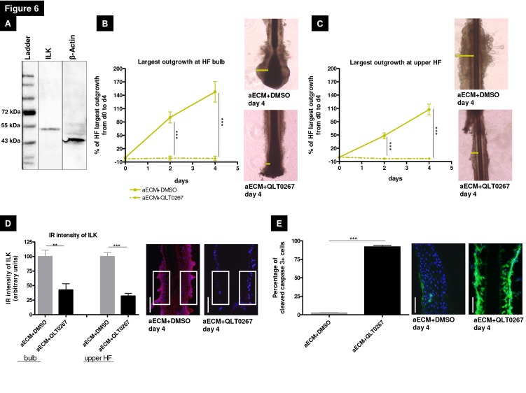
β1 integrin regulates multiple epithelial cell functions by connecting cells with the extracellular matrix (ECM). While β1 integrin-mediated signaling in murine epithelial stem cells is well-studied, its role in human adult epithelial progenitor cells (ePCs) in situ remains to be defined. Using microdissected, organ-cultured human scalp hair follicles (HFs) as a clinically relevant model for studying human ePCs within their natural topobiological habitat, β1 integrin-mediated signaling in ePC biology was explored by β1 integrin siRNA silencing, specific β1 integrin-binding antibodies and pharmacological inhibition of integrin-linked kinase (ILK), a key component of the integrin-induced signaling cascade. β1 integrin knock down reduced keratin 15 (K15) expression as well as the proliferation of outer root sheath keratinocytes (ORSKs). Embedding of HF epithelium into an ECM rich in β1 integrin ligands that mimic the HF mesenchyme significantly enhanced proliferation and migration of ORSKs, while K15 and CD200 gene and protein expression were inhibited. Employing ECM-embedded β1 integrin-activating or -inhibiting antibodies allowed to identify functionally distinct human ePC subpopulations in different compartments of the HF epithelium. The β1 integrin-inhibitory antibody reduced β1 integrin expression in situ and selectively enhanced proliferation of bulge ePCs, while the β1 integrin-stimulating antibody decreased hair matrix keratinocyte apoptosis and enhanced transferrin receptor (CD71) immunoreactivity, a marker of transit amplifying cells, but did not affect bulge ePC proliferation. That the putative ILK inhibitor QLT0267 significantly reduced ORSK migration and proliferation and induced massive ORSK apoptosis suggests a key role for ILK in mediating the ß1 integrin effects. Taken together, these findings demonstrate that ePCs in human HFs require β1 integrin-mediated signaling for survival, adhesion, and migration, and that different human HF ePC subpopulations differ in their response to β1 integrin signaling. These insights may be exploited for cell-based regenerative medicine strategies that employ human HF-derived ePCs.


Conflict of interest statement

Competing Interests: The authors have declared that no competing interests exist.

Figures

Figure 1
Figure 1. Gene silencing of β1 integrin in normal human hair follicles.
(A) ß1 integrin gene expression was analysed with qRT-PCR using full-length hair follicles (HFs). At day1 the silencing had a strong influence on the ß1 integrin transcription of HFs, PCR results on day 4 confirmed a significant silencing. Fold expression of all analyzed genes were normalized to GAPDH. n =2-3 individuals (for RNA extraction 12 HFs/patient were used and cultured over 4 days). (B) The immunoreactivity (IR) pattern of ß1 integrin in the whole HF was analyzed on day4 using the ß1 integrin-activating antibody 12G10. The IR intensity displayed no differences in the analyzed groups and in the different HF compartments. The control is normalized to 100%. n=17-26 HFs of 3 individuals; representative photos of HF bulbs on day4. (C) β1 integrin silencing caused a significant reduction of Ki-67+ matrix keratinocytes of anagen HFs (counted below Auber’s line; dotted white line) treated with β1 integrin siRNA compared to the scrambled control on day4. n=13-16 HFs of 3 individuals. (D) To dissect the proliferation capacity of slow-cycling epithelial progenitor cells of the HF bulge Ki-67+ cells were counted in rectangles (representative photos). β1 integrin silencing caused a significant reduction of Ki-67+ cells in the bulb, but also in the HF bulge on day4. n=13-24 HFs of 3 individuals. Red bars=Ki-67, green bars=TUNEL. (E) By analyzing the proliferating status in a human HF via counting EdU+ cells in our rectangles we could show the same tendency for proliferation in β1 integrin-mediated signaling as counting Ki-67+ cells in the analyzed HF compartments. n=2-3 HFs of 1 individual. White scale bars represent=50µm. All statistical analyses were performed with the one-way ANOVA by appropriate post hoc comparison (depending on a given Gaussian distribution), mean of normalized data +/- SEM (*p<0.05, **p<0.01, ***p<0.001). Abbreviation: HKG=housekeeping gene, GAPDH=glyceraldehyde3-phosphate dehydrogenase, ITGβ1 KD=knock down of ß1 integrin, IR=immunoreactivity.
Figure 2
Figure 2. β1 integrin silencing reduced significantly the epithelial progenitor protein expression in the hair follicle bulge.
(A) Keratin 15 (K15) transcription was significantly reduced at day 4 by β1 integrin siRNA compared to the scrambled control. (B) K15 immunoreactivity (IR) was most downregulated in β1 integrin siRNA silenced HFs in the upper HF (including the bulge region), n=19-26 HFs of 3 individuals. (B1-3) Representative photos demonstrating the reference areas in the upper HF. (C) β1 integrin silencing slightly enhanced the gene expression of CD200. (D) CD200 IR in the HF bulge was significantly reduced compared to the scrambled control at day 4; n=17-28 HFs of 3 individuals. (D1-3) Representative photos which show the reference areas in the upper HF. (E) β1 integrin knock down initially enhanced expression of K6, but reduced the transcription level subsequently at day 4. (F) IR intensity of K6 in the different HF compartments. It demonstrated a non-specific repression of K6 IR in every analyzed HF compartment by the silencing procedure as such, but not specifically by β1 integrin silencing. n=16-18 HFs of 2 individuals. (F1-3) Representative photos which show the reference areas in the upper HF. Fold expression of all analyzed genes were normalized to GAPDH. n=2-3 individuals (for RNA extraction 12 HFs/patient were used and cultured over 4 days). IR intensity of the HF bulb and the upper HF was measured with a specified rectangle with ImageJ (250x125). White scale bars represent=100µm. All statistical analyses were performed with the one-way ANOVA by appropriate post hoc comparison (depending on a given Gaussian distribution), mean of normalized data +/- SEM (*p<0.05, **p<0.01, ***p<0.001). Abbreviation: HKG=housekeeping gene, GAPDH=glyceraldehyde 3-phosphate dehydrogenase, ITGβ1 KD=knock down of ß1 integrin, K=keratin, IR=immunoreactivity.
Figure 3
Figure 3. β1 integrin receptor ligands differentially regulate vitality of keratinocytes in different hair follicle compartments.
(A) Epithelial outgrowth area of outer root sheath keratinocytes (ORSKs) over 4 days was measured. While the vehicle control hair follicles (HFs) showed no ORSK outgrowth in the culture dishes, the embedded HFs (aECM) showed a 30% larger ORSK outgrowth area. Activating and inhibiting β1 integrin antibodies had very similar stimulatory effects on ORSK outgrowth area. n=20-41 HFs of 3-4 individuals. Green lines and stars mark the significances of day2; black lines and stars mark the significances of day4. Statistical analysis was performed with the Mann-Whitney test; mean+/-SEM (*p<0.05, **p<0.01, ***p<0.001). (B) Representative photos of dispase-treated, embedded HF (aECM). Dotted line demarcates the area of analysis for the ORSK outgrowth over the culture period of 4 days. Black scale bar=100µm. (C) Ki-67/TUNEL-staining demonstrated that β1 integrin ligands, like extracellular matrix components and the specific receptor antibodies, decreased apoptosis in the upper HF, whereas in the aECM and aECM+mAb13-treated group the proliferation rate is up-regulated, aECM+12G10 is similar to the vehicle control in the HF bulb. n=7-15 HFs of 2-3 individuals. (D) Ki-67/TUNEL-staining confirmed the influence of β1 integrin ligands on HF bulb cells. In the aECM and aECM+12G10-treated group the number of proliferative cells in the human HF bulb significantly increased. The inhibiting antibody mAb13 enlarged apoptosis in HF bulb cells. n=8-16 HFs of 2-3 individuals. Statistical analyses of Ki-67/TUNEL were performed with the one-way ANOVA by appropriate post hoc comparison (depending on a given Gaussian distribution), error bars=mean of normalized data +/- SEM (*p<0.05, **p<0.01, ***p<0.001). Red bars=Ki-67, green bars=TUNEL. (E) Cortactin revealed activated migration mainly in the HF bulb of the aECM-treated group. White scale bars=50µm. Abbreviation: aECM=artificial ECM consisting of Matrigel®, collagen I and K-SFM (keratinocyte-serum free medium), aECM+12G10=aECM supplemented with the activating β1 integrin antibody 12G10, aECM+mAb13=aECM supplemented with the inhibiting β1 integrin antibody mAb13.
Figure 4
Figure 4. Schematic drawing for differential regulation via β1 integrin antibodies.
Comparison of the influences of activated and inhibited signaling via β1 integrin specific antibodies on the protein expression of different immunoreactivity markers. The immunoreactivity analyses of the β1 integrin-activating (12G10) or –inhibiting (mAb13) antibody-treated and aECM (artificial extracellular matrix medium) embedded hair follicle (HF) epithelium suggested a different response of the epithelial progenitors cell subpopulations on ß1 integrin signaling. The application of aECM-incorporated β1 integrin antibodies allowed distinguishing adult human epithelial progenitor cell subpopulations with distinct amplifying capacities in situ, which are located in separate epithelial compartments of human scalp HFs.
Figure 5
Figure 5. β1 integrin-mediated signaling stimulated different epithelial progenitor cell populations in distinct hair follicle compartments.
(A) Relative gene expression of β1 integrin. The β1 integrin-activating antibody 12G10 did not alter β1 integrin gene expression, whereas the inhibition of the receptor via mAb13 significantly reduced β1 integrin expression. n=1-2 individuals in experimental triplicates (15 hair follicles [HFs]). (B) Immunoreactivity of CD71 enabled to distinguish between different epithelial progenitor cell populations within the HF bulge and the HF bulb. n=5-10 HFs of 3-4 individuals. The data were analysed by using the one-way ANOVA with the appropriate post hoc comparison (depending on a given Gaussian distribution), mean of normalized data +/- SEM (*p<0.05, **p<0.01, ***p<0.001). (C) Measurement of the largest outgrowth in the upper HF and (D) HF bulb over 4 days displayed the large influence of the β1 integrin antibody treatment and distinguished between the different epithelial progenitor cell populations via their response to β1 integrin antibody stimulation. In the upper HF region (including the bulge) the inhibiting β1 integrin antibody mAb13 significantly stimulated epithelial outgrowth, whereas in the HF bulb the activating antibody 12G10 antibody stimulated epithelial outgrowth. Photos show dispase-pretreated upper HFs and HF bulbs after embedding into the aECM (artificial extracellular matrix) system and treating with β1 integrin antibodies at day 4. White lines demarcate the reference areas. n=18-33 HFs of 4 individuals. Mean+/-SEM, using unpaired t-test (*p<0.05, **p<0.01, ***p<0.001). Scale bars: 100µm. Abbreviation: aECM=artificial ECM consisting of Matrigel®, collagen I and K-SFM (keratinocyte-serum free medium), aECM+12G10=aECM supplemented with the activating β1 integrin antibody 12G10, aECM+mAb13=aECM supplemented with the inhibiting β1 integrin antibody mAb13, HKG=housekeeping gene, PPIA=peptidylprolyl isomerase A, IR = immunoreactivity.
Figure 6
Figure 6. Integrin-linked kinase inhibition via QLT0267 stops the epithelial outgrowth.
(A) Integrin-linked kinase (ILK) is expressed in human dispase-pretreated HF (hair follicle) keratinocytes, which was demonstrated by using the Western blot method in comparison to the protein expression of β-actin. (B) The analysis of the largest outgrowth in the HF bulb and (C) in the upper HF revealed the strong inhibitory effect of the pharmacological substance QLT0267 for the proliferative and migrative capacity. Representative photos. n=24-28 HFs of 3 individuals. (D) In the HF bulb and in the upper HF a significant reduction of ILK immunoreactivity could be demonstrated with QLT0267 treatment in comparison to our control HFs, which were dispase-pretreated and embedded in the artificial extracellular matrix with DMSO. Representative photos show the reference areas in the upper HF, n=7-10 HFs of 3 individuals. (E) The QLT0267 treatment caused a tremendous apoptotic effect in the HF epithelium, showing by nearly 100% cleaved caspase 3+ outer root sheath keratinocytes. n=5-10 HFs of 3 individuals. For each immunoreactivity intensity analysis the control was normalized to 100%. White scale bars=50µm. All statistical analyses were done by using Mann-Whitney test, (*p<0.05, **p < 0.01, ***p < 0.001); mean+/-SEM. Abbreviation: aECM+DMSO=artificial ECM consisting of Matrigel®, collagen I and K-SFM (keratinocyte-serum free medium), aECM+QLT0267=aECM supplemented with the 100µM pharmacological inhibitor QLT0267, DMSO=Dimethyl sulfoxide, ILK=integrin-linked kinase, IR=immunoreactivity.

Similar articles

Cited by

References

    1. Benoit YD, Lussier C, Ducharme PA, Sivret S, Schnapp LM et al. (2009) Integrin alpha8beta1 regulates adhesion, migration and proliferation of human intestinal crypt cells via a predominant RhoA/ROCK-dependent mechanism. Biol Cell 101: 695-708. doi:10.1042/BC20090060. PubMed: 19527220. - DOI - PMC - PubMed
    1. Streuli CH (2009) Integrins and cell-fate determination. J Cell Sci 122: 171-177. doi:10.1242/jcs.018945. PubMed: 19118209. - DOI - PMC - PubMed
    1. Brakebusch C, Grose R, Quondamatteo F, Ramirez A, Jorcano JL et al. (2000) Skin and hair follicle integrity is crucially dependent on beta 1 integrin expression on keratinocytes. EMBO J 19: 3990-4003. doi:10.1093/emboj/19.15.3990. PubMed: 10921880. - DOI - PMC - PubMed
    1. Hynes RO (2002) Integrins: bidirectional, allosteric signaling machines. Cell 110: 673-687. doi:10.1016/S0092-8674(02)00971-6. PubMed: 12297042. - DOI - PubMed
    1. Campbell ID, Humphries MJ (2011) Integrin structure, activation, and interactions. Cold Spring Harb Perspect Biol 3. - PMC - PubMed

Publication types

MeSH terms