. 2013 Dec 27;8(12):e84902.
doi: 10.1371/journal.pone.0084902. eCollection 2013.

Adaptation of an L-proline adenylation domain to use 4-propyl-L-proline in the evolution of lincosamide biosynthesis

Affiliations

Adaptation of an L-proline adenylation domain to use 4-propyl-L-proline in the evolution of lincosamide biosynthesis

Stanislav Kadlčík et al. PLoS One. .

Abstract

Clinically used lincosamide antibiotic lincomycin incorporates in its structure 4-propyl-L-proline (PPL), an unusual amino acid, while celesticetin, a less efficient related compound, makes use of proteinogenic L-proline. Biochemical characterization, as well as phylogenetic analysis and homology modelling combined with the molecular dynamics simulation were employed for complex comparative analysis of the orthologous protein pair LmbC and CcbC from the biosynthesis of lincomycin and celesticetin, respectively. The analysis proved the compared proteins to be the stand-alone adenylation domains strictly preferring their own natural substrate, PPL or L-proline. The LmbC substrate binding pocket is adapted to accommodate a rare PPL precursor. When compared with L-proline specific ones, several large amino acid residues were replaced by smaller ones opening a channel which allowed the alkyl side chain of PPL to be accommodated. One of the most important differences, that of the residue corresponding to V306 in CcbC changing to G308 in LmbC, was investigated in vitro and in silico. Moreover, the substrate binding pocket rearrangement also allowed LmbC to effectively adenylate 4-butyl-L-proline and 4-pentyl-L-proline, substrates with even longer alkyl side chains, producing more potent lincosamides. A shift of LmbC substrate specificity appears to be an integral part of biosynthetic pathway adaptation to the PPL acquisition. A set of genes presumably coding for the PPL biosynthesis is present in the lincomycin--but not in the celesticetin cluster; their homologs are found in biosynthetic clusters of some pyrrolobenzodiazepines (PBD) and hormaomycin. Whereas in the PBD and hormaomycin pathways the arising precursors are condensed to another amino acid moiety, the LmbC protein is the first functionally proved part of a unique condensation enzyme connecting PPL to the specialized amino sugar building unit.


Conflict of interest statement

Competing Interests: The authors have declared that no competing interests exist.

Figures

Figure 1
Figure 1. Structures of lincosamides (A–C) and other natural compounds containing branched L-proline precursors (D–G).
(A) Lincomycin A, (B) Lincomycin B, (C) Celesticetin, (D) Anthramycin, (E) Sibiromycin, (F) Tomaymycin, (G) Hormaomycin. Fragments derived from L-proline are highlighted.
Figure 2
Figure 2. Phylogenetic relationships of NRPS A-domains specific for L-proline or its derivatives.
A rooted, neighbor-joining phylogenetic tree was constructed based on the full length amino acid sequences of stand-alone A-domains and excised sequences of modular A-domains. Bootstrap values (100 replicates) above 50% are indicated at the nodes. Modular A-domains are represented by name of respective NRPS module and marked with a ■, stand-alone A-domains are marked with a ▲. The substrates for each domain include L-proline (Pro), L-proline derivatives with one carbon side chains (Pro1C), L-proline derivatives with two carbon side chains (Pro2C), and L-proline derivatives with three carbon side chain (Pro3C). Those A-domains specific for Pro2C or Pro3C substrates are highlighted. Number in parentheses behind the name of respective NRPS denotes the number of the module in NRPS protein chain, if relevant; letter in parentheses denotes the source organism. The GenBank accession numbers, producing strains, and references describing each A-domain are listed. Percent sequence identities with LmbC and CcbC were calculated from pairwise alignments. Figure S1A shows an identical phylogenetic tree reflecting phylogenetic distances, Figure S1B shows the identical set analysis using maximum likelihood method.
Figure 3
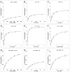
Figure 3. Comparison of the CcbC, LmbC and LmbC G308V reaction kinetics for various substrates.
The following combinations of proteins and substrates were tested: (A) LmbC vs. L-proline, (B) LmbC vs. EPL, (C) LmbC vs. PPL, (D) LmbC vs. BuPL, (E) LmbC vs. PePL, (F) CcbC vs. L-proline, (G) LmbC G308V vs. L-proline, (H) LmbC G308V vs. EPL and (I) LmbC G308V vs. PPL. All reactions were performed in triplicate. The error bars indicate the standard deviation. The reaction velocity is expressed as the amount of radioactive ATP (μM) produced per minute at protein concentration 0.05 μM. Reaction conditions are described in Experimental Section.
Figure 4
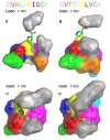
Figure 4. Homology models of the CcbC and LmbC binding pocket with the substrate.
The models of LmbC (A) and CcbC (B) at frame 805 (time 8.05 ns) of a 20-ns-long, non-restrained MD simulation. Pictures C and D at frame 788 (7.88 ns) and frame 805 (time 8.05 ns) represent another perspective of the CcbC homology model. The letters of the nonribosomal code at upper edge are colored to correspond to the individual amino acid residues of the structures.
Figure 5
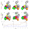
Figure 5. Homology models of the LmbC and LmbC G308V amino acid binding pocket and an RMSD analysis of these models during MD simulations.
Structures of the substrate binding pockets from LmbC (A–C) and LmbC G308V (D–F) homology models with bound PPL during the course of a 20-ns-long, non-restrained MD simulation are shown at 0 ns (left column), 8.05 ns (middle column), and 19.09 ns (right column). The nonribosomal code of each model is displayed at left. The individual letters of the code are colored to correspond to those of the individual amino acids in the structures. A time-based RMSD analysis of the substrate during a 20-ns-long, non-restrained MD simulation of LmbC (blue line) and LmbC G308V (red line). The RMSD was calculated over all substrate C atoms. The positions of the frames 0, 805 and 1909 (corresponding to the time 0 ns, 8.05 ns, and 19.09 ns) are marked with vertical lines.

References

    1. Brahme NM, Gonzalez JE, Rolls JP, Hessler EJ, Mizsak S et al. (1984) Biosynthesis of the lincomycins. 1. Studies using stable isotopes on the biosynthesis of the propyl-L-hygric and ethyl-L-hygric acid moieties of lincomycin-A and lincomycin-B. J Am Chem. Soc 106: 7873-7878.
    1. Neusser D, Schmidt H, Spizèk J, Novotnà J, Peschke U et al. (1998) The genes lmbB1 and lmbB2 of Streptomyces lincolnensis encode enzymes involved in the conversion of L-tyrosine to propylproline during the biosynthesis of the antibiotic lincomycin A. Arch Microbiol 169: 322-332. doi:10.1007/s002030050578. PubMed: 9531633. - DOI - PubMed
    1. Novotná J, Honzátko A, Bednář P, Kopecký J, Janata J et al. (2004) L-3,4-Dihydroxyphenyl alanine-extradiol cleavage is followed by intramolecular cyclization in lincomycin biosynthesis. Eur J Biochem 271: 3678-3683. doi:10.1111/j.1432-1033.2004.04308.x. PubMed: 15355345. - DOI - PubMed
    1. Hu Y, Phelan V, Ntai I, Farnet CM, Zazopoulos E et al. (2007) Benzodiazepine biosynthesis in Streptomyces refuineus . Chem Biol 14: 691-701. doi:10.1016/j.chembiol.2007.05.009. PubMed: 17584616. - DOI - PubMed
    1. Li W, Khullar A, Chou S, Sacramo A, Gerratana B (2009) Biosynthesis of sibiromycin, a potent antitumor antibiotic. Appl Environ Microbiol 75: 2869-2878. doi:10.1128/AEM.02326-08. PubMed: 19270142. - DOI - PMC - PubMed

Publication types

MeSH terms