. 2014 Jan 3;7(1):2.
doi: 10.1186/1754-6834-7-2.

Ligninolytic peroxidase genes in the oyster mushroom genome: heterologous expression, molecular structure, catalytic and stability properties, and lignin-degrading ability

Affiliations

Ligninolytic peroxidase genes in the oyster mushroom genome: heterologous expression, molecular structure, catalytic and stability properties, and lignin-degrading ability

Elena Fernández-Fueyo et al. Biotechnol Biofuels. .

Abstract

Background: The genome of Pleurotus ostreatus, an important edible mushroom and a model ligninolytic organism of interest in lignocellulose biorefineries due to its ability to delignify agricultural wastes, was sequenced with the purpose of identifying and characterizing the enzymes responsible for lignin degradation.

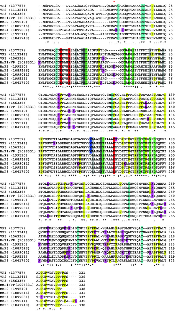
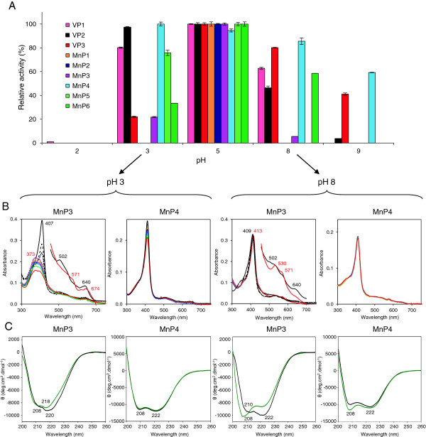
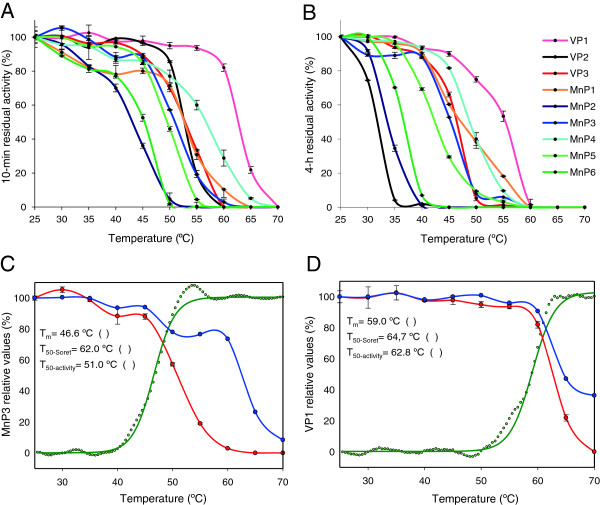
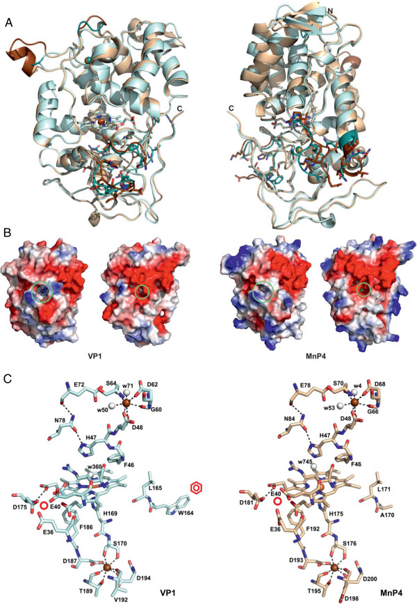
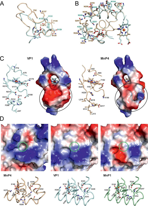
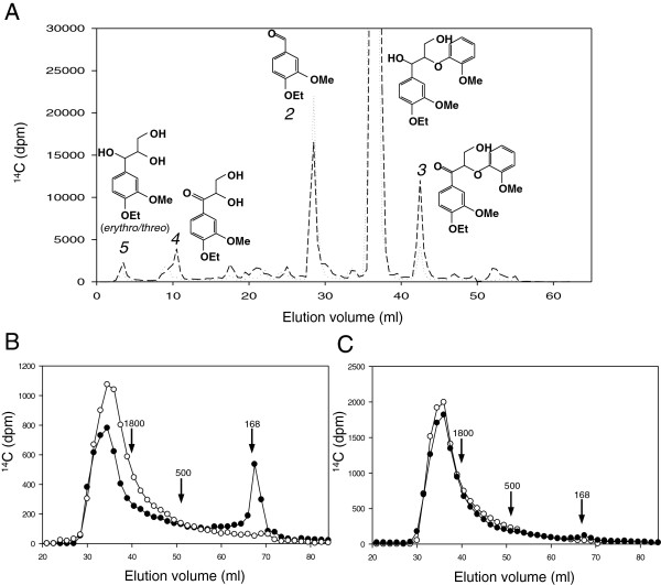
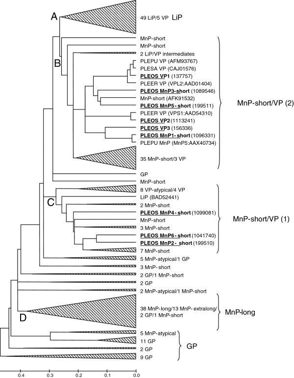
Results: Heterologous expression of the class II peroxidase genes, followed by kinetic studies, enabled their functional classification. The resulting inventory revealed the absence of lignin peroxidases (LiPs) and the presence of three versatile peroxidases (VPs) and six manganese peroxidases (MnPs), the crystal structures of two of them (VP1 and MnP4) were solved at 1.0 to 1.1 Å showing significant structural differences. Gene expansion supports the importance of both peroxidase types in the white-rot lifestyle of this fungus. Using a lignin model dimer and synthetic lignin, we showed that VP is able to degrade lignin. Moreover, the dual Mn-mediated and Mn-independent activity of P. ostreatus MnPs justifies their inclusion in a new peroxidase subfamily. The availability of the whole POD repertoire enabled investigation, at a biochemical level, of the existence of duplicated genes. Differences between isoenzymes are not limited to their kinetic constants. Surprising differences in their activity T50 and residual activity at both acidic and alkaline pH were observed. Directed mutagenesis and spectroscopic/structural information were combined to explain the catalytic and stability properties of the most interesting isoenzymes, and their evolutionary history was analyzed in the context of over 200 basidiomycete peroxidase sequences.

Conclusions: The analysis of the P. ostreatus genome shows a lignin-degrading system where the role generally played by LiP has been assumed by VP. Moreover, it enabled the first characterization of the complete set of peroxidase isoenzymes in a basidiomycete, revealing strong differences in stability properties and providing enzymes of biotechnological interest.


Figures

Figure 1
Figure 1
Multiple alignment of amino acid sequences of the nine PODs from the P. ostreatus genome (current JGI references, after manual annotation, included). Conserved catalytic and other relevant residues are indicated with different colors including: eight cysteines (cyan) forming four disulfide bridges; nine ligands (green) of two structural Ca2+ ions; two active site histidines (dark gray); three acidic residues (orange) forming the Mn2+ oxidation site; one tryptophan (blue) responsible for aromatic substrate oxidation by VPs (also present in MnP1, initially classified as a VP); several active site conserved residues (light gray); and several lysine (purple) and proline (yellow) residues being particularly frequent in some of the sequences. Alignment was prepared using ClustalW2 (European Bioinformatics Institute, Hinxton, UK). Amino acid numbering starts at the first residue of the mature protein (red asterisk on the alignment). Symbols below indicate full conservation of the same (*) or equivalent residues (:) and partial residue conservation (.). JGI, Joint Genome Institute; MnP, manganese peroxidase; POD, class II peroxidase from the superfamily of non-animal (plant-fungal-prokaryotic) peroxidases; VP, versatile peroxidase.
Figure 2
Figure 2
pH stabilities of the nine PODs from the P. ostreatus genome. (A) Residual activities after 4 hours of incubation at five selected pH values in the pH 2 to 9 range. Residual activities of the E. coli-expressed and in vitro-activated three VP and six MnP isoenzymes were determined (by 5 mM ABTS oxidation in 0.1 M tartrate, pH 3.5, except for MnP1 and MnP6, for which 2 mM ABTS was used) after incubation (at 4°C) in 100 mM B&R buffer of pH 2 to 9 (Additional file 1: Figure S4 shows data at eight pH values after 1 minute, 1, 4, 24, and 120 hours of incubation, and Figure S5 shows comparison of pH stability at 4°C and 25°C). Means and 95% confidence limits. (B) UV-visible spectra of unstable MnP3 and stable MnP4 isoenzymes after 0 (black), 30 (magenta), 60 (yellow), 90 (purple), 120 (blue), 180 (green), and 240 minutes (red) of incubation at pH 3 and 8. Three additional scans at 30 (large dashes), 60 (short dashes), and 90 seconds (dots) of incubation, and amplified (x 5 absorbance) 450 to 700 nm region are shown for MnP3 initial and final spectra. Main maxima are indicated. (C) Far-UV CD spectra of unstable MnP3 and stable MnP4 isoenzymes after 1 minute (black) and 1 hour (green) of incubation at pH 3 and 5. Results are shown as molar ellipticities (θ) and main minima are indicated. All spectra were recorded at 25°C. ABTS, 2,2′-azino-bis(3-ethylbenzothiazoline-6-sulfonate); B&R, Britton-Robinson; CD, circular dichroism; MnP, manganese peroxidase; POD, class II peroxidase from the superfamily of non-animal (plant-fungal-prokaryotic) peroxidases; VP, versatile peroxidase.
Figure 3
Figure 3
Thermal stabilities of the nine PODs from the P. ostreatus genome. (A, B) Residual activities after 10 minutes and 4 hours of incubation, respectively, in the range of 25 to 70°C. Residual activities were determined as described in Figure 2 after 10 minutes and 4 hours of incubation in 10 mM tartrate (pH 5) at ten temperatures (5°C intervals). Means and 95% confidence limits. From the above curves, the 10-minute and 4-hour T50-activity values were obtained (Additional file 2: Table S2). (C, D) Denaturation of temperature-unstable MnP3 and stable VP1, respectively, as shown by UV-visible (blue) and CD (green) spectroscopy, compared with activity lost (red). The effect of temperature (25 to 70°C) is shown by the θ increase at 222 nm in CD spectra acquired each 0.5°C, the absorbance decrease at the Soret maximum (at 407 nm) in the UV-visible spectra acquired each 5°C, and the decrease of enzyme residual activity after 10 minutes of incubation, estimated as described in Figure 2. All measurements were performed in 10 mM tartrate (pH 5). T50 values corresponding to those temperatures where 50% protein denaturation (main melting transition) is shown by CD spectra (Tm), 50% decrease of heme Soret band is shown by UV-visible spectra (T50-Soret), and 50% decrease of enzymatic activity is produced (T50-activity; means and 95% confidence limits) are included. CD, circular dichroism; MnP, manganese peroxidase; POD, class II peroxidase from the superfamily of non-animal (plant-fungal-prokaryotic) peroxidases; VP, versatile peroxidase.
Figure 4
Figure 4
Crystal structures of the most interesting VP/MnP isoenzymes from the P. ostreatus genome (1.05 to 1.10 Å). (A) Superposition of the overall structures of VP1 (light blue) and MnP4 (light brown) from two different orientations. The significant differences between the structures are shown in darker color with some side chains of these regions as sticks. (B) Electrostatic surface of VP1 and MnP4 from two different orientations showing the main heme access channel (larger circle) and the narrower Mn2+ access channel (smaller circle) as well as the more basic MnP4 surface with a high number of exposed lysine residues. (C) Detail of the VP1 and MnP4 heme and neighbor regions showing: proximal histidine (H169 and H175, respectively); distal histidine (H47) and neighbor arginine (R43); two conserved phenylalanines at the proximal histidine side (F186 and F182, respectively) and the distal histidine side (F46) of the heme; two conserved asparagines (N78 and N84, respectively) and glutamate residues (E72 and E78, respectively) forming a H-bond network from the distal histidine; two structural Ca2+ ions (brown spheres) with their four/five conserved ligands; one site for oxidation of Mn2+ (whose predicted position is marked with a red circle) near the internal propionate of heme, formed by two glutamates (E36 and E40) and one aspartate (D175 and D181, respectively); one tryptophan residue (W164) constituting the site (marked with a red benzenic ring) of VP1 oxidation of aromatic substrates by LRET via a contiguous leucine residue (L165); and several water molecules (white spheres) near the distal histidine and Ca2+ ion. From entries [PDB:4BLK] (VP1) and [PDB:4BM1] (MnP4). LRET, long-range electron transfer; MnP, manganese peroxidase; PDB, Protein Data Bank; VP, versatile peroxidase.
Figure 5
Figure 5
Comparison of three regions in the VP1 (blue) and MnP4 (brown) crystal structures; and catalytic tryptophan environment in VP1 and homologous regions in MnP4 and MnP1. (A) Loop close to the distal calcium and its ligands including two water molecules (light spheres) and differences in the VP1/MnP4 D129 to A132/N134 to K137 region including MnP4 K137 interacting with the F62 side chain. (B) Two sequence stretches (V248 to P252 and P286 to H293 in VP1, and I254 to S259 and R292 to P298 in MnP4) at the back of the protein with indication of those residues differing in the two peroxidases (the heme cofactor, proximal Ca2+, spheres, and its ligands, and VP1 W164 are also visible). (C) Selected residues and solvent access surface in the VP1 and MnP4 heme access channel and surrounding area (differences in surface electronegativity below the channel entrance are indicated with circles). (D) Structure (bottom) and electrostatic surface (top) showing the catalytic tryptophan and surrounding residues of VP1 (including G260 and F197, among others) compared with the same region in MnP1 (including D261 and I198) and MnP4 (including R266 and F203) (the green circles represent the position of the conserved tryptophan in VP1 and MnP1, or the corresponding alanine in MnP4). From entries [PDB:4BM1] (MnP4) and [PDB:4BLK] (VP1), and MnP1 homology model based on [PDB:4BLK]. MnP, manganese peroxidase; PDB, Protein Data Bank; VP, versatile peroxidase.
Figure 6
Figure 6
Demonstration of lignin-degrading ability of P. ostreatus VP. (A) Oxidative degradation of a nonphenolic lignin model dimer by VP1. The erythro (dotted line) and threo (dashed line) isomers of 14C-labeled 4-ethoxy-3-methoxyphenylglycerol-β-guaiacyl ether (peak 1) were treated with VP1, and the completed reactions were analyzed by HPLC. The main products were 4-ethoxy-3-methoxybenzaldehyde (peak 2) and 1-(4-ethoxy-3-methoxyphenyl)-3-hydroxy-2-(2-methoxyphenoxy)-propan-1-one (peak 3). The minor peaks 4 and 5 correspond to 1-(4-ethoxy-3-methoxyphenyl)-2,3-dihydroxypropan-1-one and 1-(4-ethoxy-3-methoxyphenyl)glycerol, respectively. (B, C) Lignin depolymerization by VP1.14C-labeled synthetic lignin (DHP) was treated with VP1 from the P. ostreatus genome in the presence (B) and absence (C) of VA. Black symbols indicate reactions with enzyme and open symbols indicate controls without enzyme. Total recoveries of initially added 14C from complete reactions before GPC analysis were 62% for reaction B and 92% for reaction C. Recoveries from control reactions were somewhat higher as reported earlier [22]. The arrows indicate elution volumes of two polystyrene molecular mass standards (1,800 and 500 Da) and VA (168 Da). DHP, dehydrogenation polymer; GPC, gel permeation chromatography; HPLC, high performance liquid chromatography; VA, veratryl alcohol; VP, versatile peroxidase.
Figure 7
Figure 7
Phylogram of 237 sequences of basidiomycete PODs including the nine sequences from P. ostreatus genome (underlined). POD sequences from 21 genomes (and GenBank) were analyzed. Four main clusters were identified corresponding to LiP (A), MnP-short/VP clusters 1 and 2 (B, C), and MnP-long (D), together with the most ancestral group of GPs and unclustered sequences. Those clusters/subclusters where Pleurotus (PLEOS, P. ostreatus; PLEER, P. eryngii; PLEPU, P. pulmonarius; and PLESA, P. sapidus) sequences are not included were collapsed, with indication of the number and type of sequences included. Two LiP-type enzymes from G. subvermispora representing LiP/VP transition stages [47] are indicated, as well as a Cerrena unicolor MnP (AFK91532) related to P. ostreatus short MnPs, and the unique Trametes cervina LiP (BAD52441). MnP-atypical corresponds to a MnP type with only two acidic residues at the oxidation site [16]. Protein model numbers are provided for the nine P. ostreatus genome sequences, and GenBank references for other six POD sequences. See Ruiz-Dueñas and Martínez [19] for references of other basidiomycete PODs. GP, generic peroxidase; LiP, lignin peroxidase; MnP, manganese peroxidase; POD, class II peroxidase from the superfamily of non-animal (plant-fungal-prokaryotic) peroxidases; VP, versatile peroxidase.

References

    1. Sánchez C. Cultivation of Pleurotus ostreatus and other edible mushrooms. Appl Microbiol Biotechnol. 2010;85:1321–1337. doi: 10.1007/s00253-009-2343-7. - DOI - PubMed
    1. Gunde-Cimerman N, Cimerman A. Pleurotus fruiting bodies contain the inhibitor of 3-hydroxy-3-methylglutaryl-coenzyme-A reductase-lovastatin. Exp Mycol. 1995;19:1–6. doi: 10.1006/emyc.1995.1001. - DOI - PubMed
    1. Lavi I, Levinson D, Peri I, Tekoah Y, Hadar Y, Schwartz B. Chemical characterization, antiproliferative and antiadhesive properties of polysaccharides extracted from Pleurotus pulmonarius mycelium and fruiting bodies. Appl Microbiol Biotechnol. 2010;85:1977–1990. doi: 10.1007/s00253-009-2296-x. - DOI - PubMed
    1. Ruiz-Dueñas FJ, Martínez AT. Microbial degradation of lignin: how a bulky recalcitrant polymer is efficiently recycled in nature and how we can take advantage of this. Microbial Biotechnol. 2009;2:164–177. doi: 10.1111/j.1751-7915.2008.00078.x. - DOI - PMC - PubMed
    1. Martínez AT, Camarero S, Guillén F, Gutiérrez A, Muñoz C, Varela E, Martínez MJ, Barrasa JM, Ruel K, Pelayo M. Progress in biopulping of non-woody materials: chemical, enzymatic and ultrastructural aspects of wheat-straw delignification with ligninolytic fungi from the genus Pleurotus. FEMS Microbiol Rev. 1994;13:265–274.