

An effective approach to prevent immune rejection of human ESC-derived allografts
- PMID: 24388175
- PMCID: PMC4023958
- DOI: 10.1016/j.stem.2013.11.014
An effective approach to prevent immune rejection of human ESC-derived allografts
Abstract
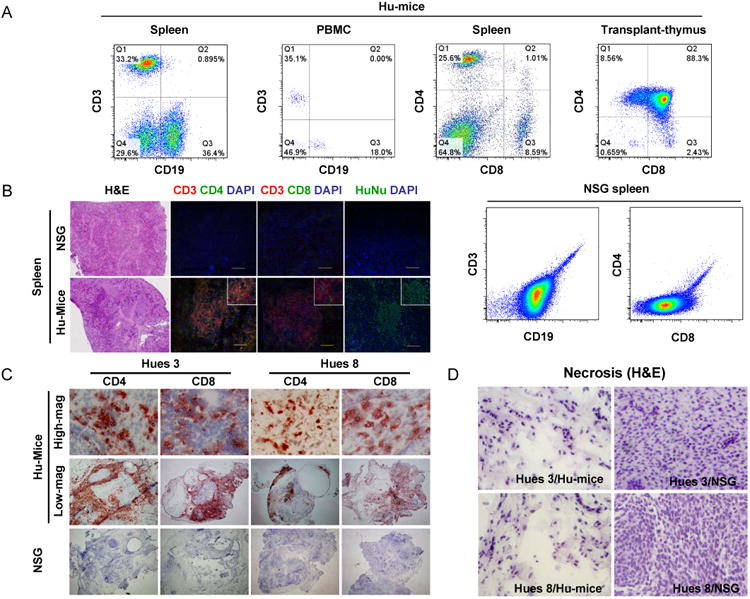
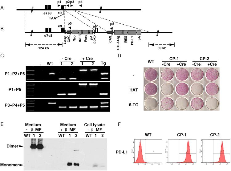
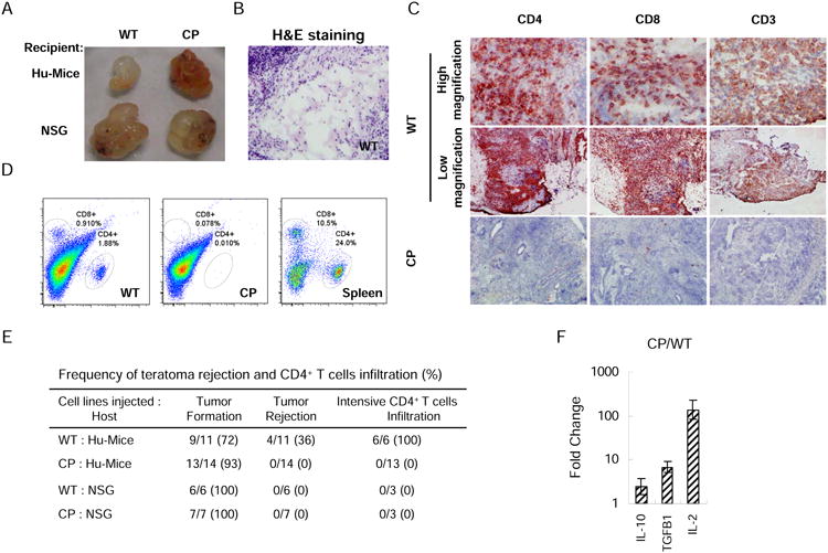
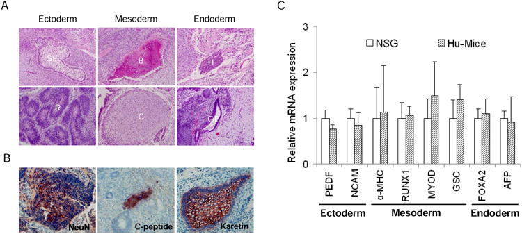
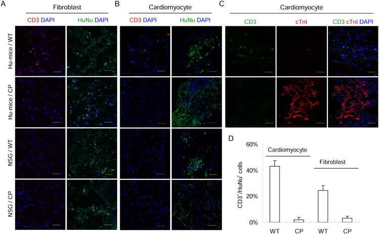
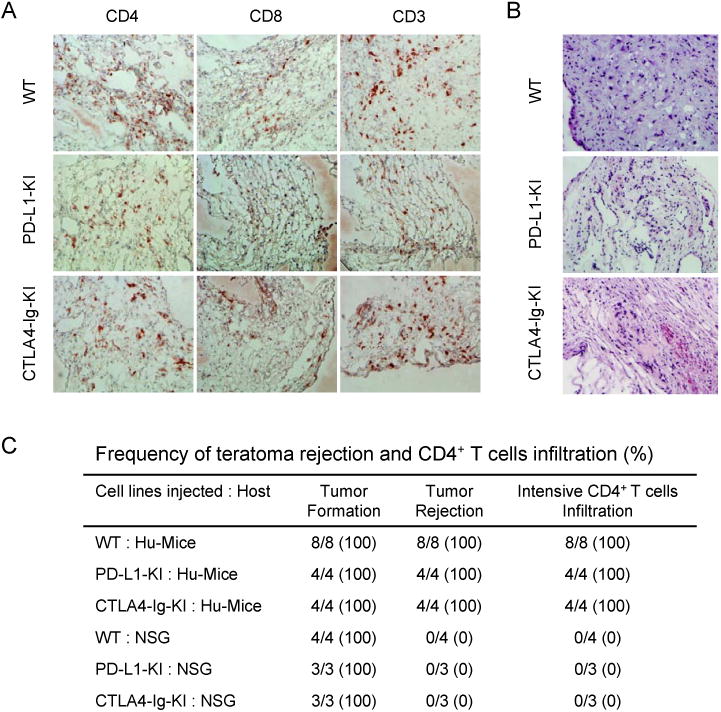
Human embryonic stem cells (hESCs) hold great promise for cell therapy as a source of diverse differentiated cell types. One key bottleneck to realizing such potential is allogenic immune rejection of hESC-derived cells by recipients. Here, we optimized humanized mice (Hu-mice) reconstituted with a functional human immune system that mounts a vigorous rejection of hESCs and their derivatives. We established knockin hESCs that constitutively express CTLA4-Ig and PD-L1 before and after differentiation, denoted CP hESCs. We then demonstrated that allogenic CP hESC-derived teratomas, fibroblasts, and cardiomyocytes are immune protected in Hu-mice, while cells derived from parental hESCs are effectively rejected. Expression of both CTLA4-Ig, which disrupts T cell costimulatory pathways, and PD-L1, which activates T cell inhibitory pathway, is required to confer immune protection, as neither was sufficient on their own. These findings are instrumental for developing a strategy to protect hESC-derived cells from allogenic immune responses without requiring systemic immune suppression.
Copyright © 2014 Elsevier Inc. All rights reserved.
Conflict of interest statement
The authors declare no conflict of interest.
Figures
Comment in
-
ESCaping rejection: A step forward for embryonic-stem-cell-based regenerative medicine.Cell Stem Cell. 2014 Jan 2;14(1):3-4. doi: 10.1016/j.stem.2013.12.003. Cell Stem Cell. 2014. PMID: 24388169
Similar articles
-
A Safety Checkpoint to Eliminate Cancer Risk of the Immune Evasive Cells Derived from Human Embryonic Stem Cells.Stem Cells. 2017 May;35(5):1154-1161. doi: 10.1002/stem.2568. Epub 2017 Feb 23. Stem Cells. 2017. PMID: 28090751
-
hESC-derived immune suppressive dendritic cells induce immune tolerance of parental hESC-derived allografts.EBioMedicine. 2020 Dec;62:103120. doi: 10.1016/j.ebiom.2020.103120. Epub 2020 Nov 23. EBioMedicine. 2020. PMID: 33242828 Free PMC article.
-
Costimulation-adhesion blockade is superior to cyclosporine A and prednisone immunosuppressive therapy for preventing rejection of differentiated human embryonic stem cells following transplantation.Stem Cells. 2013 Nov;31(11):2354-63. doi: 10.1002/stem.1501. Stem Cells. 2013. PMID: 24038578 Free PMC article.
-
Embryonic stem cell transplantation for the treatment of myocardial infarction: immune privilege or rejection.Transpl Immunol. 2007 Nov;18(2):88-93. doi: 10.1016/j.trim.2007.05.003. Epub 2007 Jun 8. Transpl Immunol. 2007. PMID: 18005850 Review.
-
Human embryonic stem cells: potential tool for achieving immunotolerance?Stem Cell Rev. 2005;1(2):151-8. doi: 10.1385/SCR:1:2:151. Stem Cell Rev. 2005. PMID: 17142850 Review.
Cited by
-
Cell therapy for Parkinson's disease with induced pluripotent stem cells.Inflamm Regen. 2023 Feb 27;43(1):16. doi: 10.1186/s41232-023-00269-3. Inflamm Regen. 2023. PMID: 36843101 Free PMC article. Review.
-
How Safe Are Universal Pluripotent Stem Cells?Cell Stem Cell. 2020 Mar 5;26(3):307-308. doi: 10.1016/j.stem.2020.02.006. Cell Stem Cell. 2020. PMID: 32142661 Free PMC article. No abstract available.
-
Generation of inner ear sensory neurons using blastocyst complementation in a Neurog1+/--deficient mouse.Stem Cell Res Ther. 2021 Feb 12;12(1):122. doi: 10.1186/s13287-021-02184-1. Stem Cell Res Ther. 2021. PMID: 33579352 Free PMC article.
-
Cell-Based Therapy for Canavan Disease Using Human iPSC-Derived NPCs and OPCs.Adv Sci (Weinh). 2020 Oct 29;7(23):2002155. doi: 10.1002/advs.202002155. eCollection 2020 Dec. Adv Sci (Weinh). 2020. PMID: 33304759 Free PMC article.
-
Translating cell therapies for neurodegenerative diseases: Huntington's disease as a model disorder.Brain. 2022 Jun 3;145(5):1584-1597. doi: 10.1093/brain/awac086. Brain. 2022. PMID: 35262656 Free PMC article.
References
-
- Blomer U, Gruh I, Witschel H, Haverich A, Martin U. Shuttle of lentiviral vectors via transplanted cells in vivo. Gene Ther. 2004;12:67–74. - PubMed
-
- Boyd AS, Rodrigues NP, Lui KO, Fu X, Xu Y. Concise Review: Immune Recognition of Induced Pluripotent Stem Cells. STEM CELLS. 2012;30:797–803. - PubMed
-
- Cheng F, Ke Q, Chen F, Cai B, Gao Y, Ye C, Wang D, Zhang L, Lahn BT, Li W, et al. Protecting against wayward human induced pluripotent stem cells with a suicide gene. Biomaterials. 2012;33:3195–3204. - PubMed
-
- Ciancio G, Garcia-Morales R, Mathew J, Carreno M, Burke GW, Ricordi C, Kenyon N, Esquenazi V, Cirocco R, Tzakis A, et al. Donor bone marrow infusions are tolerogenic in human renal transplantation. Transplantation Proceedings. 2001;33:1295–1296. - PubMed
-
- Cohen DE, Melton D. Turning straw into gold: directing cell fate for regenerative medicine. Nat Rev Genet. 2011;12:243–252. - PubMed
Publication types
MeSH terms
Substances
Grants and funding
LinkOut - more resources
Full Text Sources
Other Literature Sources
Research Materials
Miscellaneous
