. 2014 Mar;35(8):2401-2410.
doi: 10.1016/j.biomaterials.2013.11.037. Epub 2013 Dec 31.

Spatial control of adult stem cell fate using nanotopographic cues

Affiliations

Spatial control of adult stem cell fate using nanotopographic cues

Eun Hyun Ahn et al. Biomaterials. 2014 Mar.

Abstract

Adult stem cells hold great promise as a source of diverse terminally differentiated cell types for tissue engineering applications. However, due to the complexity of chemical and mechanical cues specifying differentiation outcomes, development of arbitrarily complex geometric and structural arrangements of cells, adopting multiple fates from the same initial stem cell population, has been difficult. Here, we show that the topography of the cell adhesion substratum can be an instructive cue to adult stem cells and topographical variations can strongly bias the differentiation outcome of the cells towards adipocyte or osteocyte fates. Switches in cell fate decision from adipogenic to osteogenic lineages were accompanied by changes in cytoskeletal stiffness, spanning a considerable range in the cell softness/rigidity spectrum. Our findings suggest that human mesenchymal stem cells (hMSC) can respond to the varying density of nanotopographical cues by regulating their internal cytoskeletal network and use these mechanical changes to guide them toward making cell fate decisions. We used this finding to design a complex two-dimensional pattern of co-localized cells preferentially adopting two alternative fates, thus paving the road for designing and building more complex tissue constructs with diverse biomedical applications.

Keywords: Adipogenesis; Capillary force lithography; Differentiation; Human mesenchymal stem cells; Nanotopography; Osteogenesis.


Figures

Fig. 1
Fig. 1
Fabrication of scalable nanopatterns with defined densities of nanoposts. (A) A schematic showing nanopatterns with varying nanopost densities fabricated using capillary force lithography (CFL). Nanoposts were created at mutual post distances (d) of 1.2, 2.4, 3.6, and 5.6 μm. (B) Scanning electron microscopy (SEM) images of nanopatterned arrays with varying post densities. All the above patterns, with flat control, were fabricated on a single glass coverslip to reduce experimental variation. A photo of the coverslip is shown in the middle. The diameter of the posts is 700 nm with the varying distances between two adjacent posts. The post-to-post distances were 1.2 (top left), 2.4 (top right), 3.6 (bottom left) and 5.6 μm (bottom right). (C; D) hMSC were cultured for 14 days in a mixture (1:1, vol: vol) of adipogenesis and osteogenesis differentiation media on flat control (without nanoposts) and substrata with varying post-to-post distances. Cell circularity (C) and cell spreading area (D) of hMSC were analyzed.
Fig. 2
Fig. 2
Nanopost density regulates hMSC fate. (A-F) hMSC were cultured for 14 days in a mixture (1:1, vol: vol) of adipogenesis and osteogenesis differentiation media on 1.2 μm (A), 2.4 μm (B), 3.6 μm (C), 5.6 μm (D) and flat control (E) surfaces and then stained with Oil Red O (adipocyte marker) and alkaline phosphatase (osteocyte marker). (F) Percentage of hMSC stained with osteogenic (Grey bars) and adipogenic markers (Black bars) are shown. (G-J) The expression of adipocyte (LPL, PPARγ) and osteocyte (ALP, RUNX2)-specific markers were determined using qRT-PCR assays in hMSC cultured on flat control (without nanoposts) and substrata with nanoposts at post-to-post distances of 1.2 μm (dense) and 5.6 μm (sparse). The mRNA expression levels of ALP, RUNX2, LPL, and PPARγ were normalized to the expression of 18S rRNA (mean ± S.D., n=3).
Fig. 3
Fig. 3
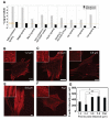
Differentially regulated pathways in osteogenesis and adipogenesis during hMSC differentiation (A) and the immunofluorescence staining of F-actin in hMSC (B-G). (A) Canonical processes that are differentially regulated in osteogenesis and adipogenesis during hMSC differentiation were analyzed using meta-analysis of published microarray data (as mentioned in Supplementary methods). Differentially expressed genes were analyzed to locate canonical biological themes that regulate cytoskeletal changes or adhesion mediated pathways. Orange line indicates the threshold (with q-value of 0.05) based on Fischer exact test. All mentioned processes are differentially expressed in datasets, suggesting that adhesion/cytoskeletal regulators are important during osteogenesis and adipogenesis. (B-G) Cytoskeletal organization of hMSC is influenced by nanopost density. hMSC were cultured for 2 days from the cell seeding on substrata of nanoposts with post-to-post distances of 1.2 μm, 2.4 μm, 3.6 μm, 5.6 μm, and flat control. (B-F) Representative images of F-actin immunofluorescence staining of hMSC exhibit a higher cortical actin organization on smaller post-to-post distance nanopatterns, while actin stress fibers are more abundant in cells cultured on larger post-to-post distance nanopatterns. A part of each photo of cells cultured on each substratum was magnified 2.5 times and shown individually in the inset. (G) The mean intensity of F-actin per cell was quantitated. Data are mean ± S.E.M [1.2 μm (n=21), 2.4 μm (n=30), 3.6 μm (n=32), 5.6 μm (n=44), and flat (n=24)].
Fig. 4
Fig. 4
Nanopost density regulates hMSC rheology correlated with observed rheological alterterations in differentiation. (A-C) Time course magnetic twisting cytometry analyses showing elastic modulus and loss modulus of hMSC cultured in the presence of osteogenic (blue circles), adipogenic (red triangles), or a 1:1 mixture (vol:vol) of osteogenic and adipogenic media (green squares) for (A) 1 day, (B) 7 days, and (C) 14 days on flat control substrata. (D-F) hMSC were cultured for 14 days in a mixed differentiation media (adipogenesis: osteogenesis, 1:1) on substrata with various post-to-post distances. Representative images (D) and elastic (E) and loss (F) moduli of hMSC on substrata with post-to-post distances of 1.2 μm (red), 2.4 μm (green), 3.6 μm (purple), and 5.6 μm (blue squares).
Fig. 5
Fig. 5
Creation of large surface area nanopost arrays to control spatial differentiation of hMSC. (A) A schematic showing the conceptual design of large surface area nanopost arrays with dense (D; 1.2 μm) and sparse (S; 5.6 μm) arrangements in a chessboard like fashion. Nanopost densities can be controlled in arbitrary fashion in two-dimensions. (B; C) hMSC were cultured for three weeks in a mixture (1:1, vol: vol) of adipogenesis and osteogenesis differentiation media on substrata with various post-to-post distances (1.2 μm; 5.6 μm) and then the cells were stained with Oil red O and alkaline phosphatase. (B) Staining images of hMSC cultured for three weeks on substrata with various post-to-post distances (1.2 μm; 5.6 μm) are shown with scanning electron microscopy (SEM) images of nanopatterns. (C) Stitched staining images of hMSC cultured for three weeks on substrata with variable post-to-post distances (1.2 μm; 5.6 μm). A higher level of Oil red O staining (an adipogenesis marker) was shown on smaller post-to-post distance areas (dense arrays), while a higher level of alkaline phosphatase staining (an osteogenesis marker) was shown on larger post-to-post distance areas (sparse arrays). The white bar indicates 50 μm. (D) Kymographic staining analysis for the percentage of adipogenic and osteogenic fate of hMSC cultured on a large surface area substratum with various post-to-post distances. Differential degrees of osteogenesis (blue) and adipogenesis (red) are shown in variations of the two colors, blue and red.

References

    1. Palpant NJ, Murry CE. Regenerative medicine: reprogramming the injured heart. Nature. 2012;485:585–6. - PubMed
    1. Delli Carri A, Onorati M, Castiglioni V, Faedo A, Camnasio S, Toselli M, et al. Human pluripotent stem cell differentiation into authentic striatal projection neurons. Stem Cell Rev. 2013;9:461–74. - PubMed
    1. Masaki T, Qu JR, Cholewa-Waclaw J, Burr K, Raaum R, Rambukkana A. Reprogramming adult schwann cells to stem cell-like cells by leprosy bacilli promotes dissemination of infection. Cell. 2013;152:51–67. - PMC - PubMed
    1. Marban E, Malliaras K. Mixed results for bone marrow-derived cell therapy for ischemic heart disease. JAMA. 2012;308:2405–6. - PubMed
    1. Makkar RR, Smith RR, Cheng K, Malliaras K, Thomson LE, Berman D, et al. Intracoronary cardiosphere-derived cells for heart regeneration after myocardial infarction (CADUCEUS): a prospective, randomised phase 1 trial. Lancet. 2012;379:895–904. - PMC - PubMed

Publication types