. 2014 Jan 16;6(1):130-40.
doi: 10.1016/j.celrep.2013.12.027. Epub 2014 Jan 2.

Genetics of follicular lymphoma transformation

Affiliations

Genetics of follicular lymphoma transformation

Laura Pasqualucci et al. Cell Rep. .

Abstract

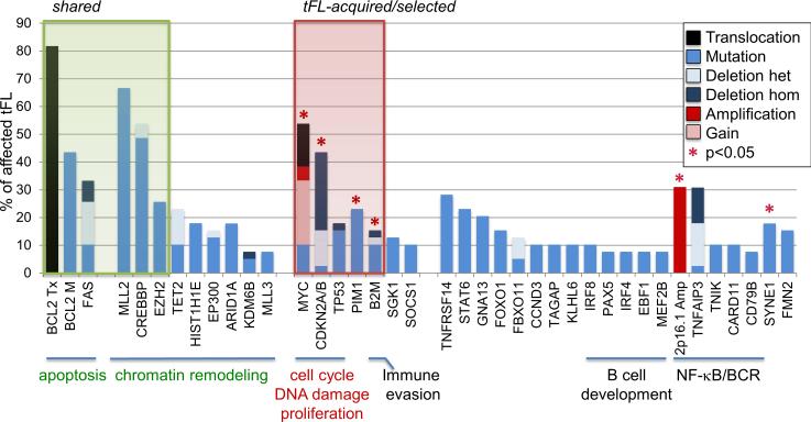
Follicular lymphoma (FL) is an indolent disease, but 30%-40% of cases undergo histologic transformation to an aggressive malignancy, typically represented by diffuse large B cell lymphoma (DLBCL). The pathogenesis of this process remains largely unknown. Using whole-exome sequencing and copy-number analysis, we show here that the dominant clone of FL and transformed FL (tFL) arise by divergent evolution from a common mutated precursor through the acquisition of distinct genetic events. Mutations in epigenetic modifiers and antiapoptotic genes are introduced early in the common precursor, whereas tFL is specifically associated with alterations deregulating cell-cycle progression and DNA damage responses (CDKN2A/B, MYC, and TP53) as well as aberrant somatic hypermutation. The genomic profile of tFL shares similarities with that of germinal center B cell-type de novo DLBCL but also displays unique combinations of altered genes with diagnostic and therapeutic implications.


Figures

Figure 1
Figure 1. FL and tFL display shared and unique genomic aberrations
Overall load of genetic lesions identified by WES and CN analysis in the dominant clone of the 12 discovery cases. Color codes denote distinct types of aberrations (Tx, translocation). *In cases lacking matched normal DNA, shared SNVs are limited to those affecting 52 selected genes with well-established roles in lymphomagenesis (see Methods); thus, the total number of genetic lesions in these patients (right column) likely represents an underestimate. FL-specific SNVs that could be due to genomic loss or cnLOH of the same region in the tFL phase were excluded.
Figure 2
Figure 2. FL and tFL arise through divergent evolution in most patients
Inferred models of clonal evolution during FL transformation. The original B cell clone is on top; blue and red circles depict the FL and tFL clones, respectively, while green, dotted circles represent the postulated common mutated precursor cell (CPC). In the linear model (2/12 patients analyzed), the tFL dominant clone originates directly from the FL dominant clone after acquisition of additional mutations. In the divergent model (10/12 patients analyzed), the FL and tFL dominant clones derive from a CPC through the independent acquisition of distinct mutations. In both scenarios, the tFL dominant clone may be present as a minor subclone already at the time of the FL biopsy. ABC, mutations shared between FL and tFL; DEF, mutations unique to the FL dominant clone; GHI, mutations unique to the tFL dominant clone.
Figure 3
Figure 3. Recurrently mutated genes in tFL
Proportion of tFL cases (n=39) carrying genetic lesions (SNVs and CNAs) in genes altered at ≥10% frequency and deemed significant by one of three independent algorithms (see Supplemental Information and Table S5); additional genes of functional relevance in the same pathways are also shown. Deletions and gains were only computed if defining MCRs of aberration encompassing maximum 3 genes. Green shade highlights alterations consistently shared between the FL and tFL sample, and red shade identifies alterations enriched in tFL (asterisks, p<0.05 when comparing tFL frequency vs unselected FL frequency; note that TP53 was considered as a tFL-specific target despite its borderline p value, because invariably unmutated in the diagnostic FL sample of all 3 informative tFL cases, and because of the broad literature data. The remaining genes did not appear to be phase-specific or could not be unequivocally assigned to a given category because of the relatively small numbers (for the full list of SNVs found in >5% of tFL cases, see Table S7).
Figure 4
Figure 4. Biallelic loss of CDKN2A/B is specifically acquired during transformation
(A) Segmentation data from 18 DLBCL samples harboring CDKN2A/B deletions, visualized using the Integrative Genomics Viewer software (http://www.broadinstitute.org/igv). Each track represents one sample, where white denotes a normal (diploid) copy number, blue indicates a region of copy number loss, and red corresponds to a region of copy number gain. The inferred copy number, and the corresponding color intensity, may vary across samples due to the presence of non-tumor cells infiltrating the biopsy. Individual genes in the region are aligned in the bottom panel, and the boxed area (defined by the red bar at the top) corresponds to the CDKN2A/B locus. (B) dChipSNP heatmap showing median-smoothed log2 CN ratio in 14 tFL biopsies harboring CDKN2A/B deletions, including 6 with matched FL biopsies, and two control DNAs (N). A vertical blue bar indicates the location of the CDKN2A/B loci. (C) Relative distribution of biallelic lesions affecting CDKN2A/B and TP53. Columns correspond to individual patients, and the genomic status of the two genes is color-coded as indicated. (D) Samples carrying biallelic alterations of CDKN2A/B and/or TP53 are characterized by a significantly higher number of CN aberrations (Mann-Whitney U test, p=0.03).
Figure 5
Figure 5. Recurrent genetic lesions of MYC in tFL
(A) GISTIC analysis of CN gains in FL and tFL cases (see also Figure S5). (B) Percentage of cases carrying genetic lesions of MYC in FL and tFL. (C) FISH analysis using a MYC break-apart probe in tFL#40. (D) IHC analysis of MYC expression in the pre- and post-transformation biopsy of the same patient.
Figure 6
Figure 6. ASHM is associated with transformation
(A) Proportion of cases harboring mutations in known ASHM targets; the BCL6 intron 1 region, a physiologic target of SHM in GC B cells, and the BCL2 sequences, which accumulate mutations in translocated alleles under the influence of the juxtaposed IGH promoter, are shown as controls. The three mutated FL cases harbored one single event each.
Figure 7
Figure 7. tFL displays a unique genomic profile that partially overlaps with GCB-DLBCL
Percentage of cases carrying the indicated genetic lesions in de novo DLBCL (top panel: green bars, GCB-DLBCL; n=50; red bars, ABC-DLBCL; n=52) and tFL (bottom panel, black bars; n=39). Asterisks indicate statistically significant differences (one-tailed Fisher's exact test; * p<0.05, ** p<0.01). Mutation frequencies for SGK1 and GNA13 are derived from Morin et al., Nature 2011. M, mutation (missense, nonsense, frameshift, splice-site); D, deletion; G, gain; AMP, amplification; M/D*, biallelic inactivation; Tx, translocation.

References

    1. Akasaka T, Lossos IS, Levy R. BCL6 gene translocation in follicular lymphoma: a harbinger of eventual transformation to diffuse aggressive lymphoma. Blood. 2003;102:1443–1448. - PubMed
    1. Alizadeh AA, Eisen MB, Davis RE, Ma C, Lossos IS, Rosenwald A, Boldrick JC, Sabet H, Tran T, Yu X, et al. Distinct types of diffuse large B-cell lymphoma identified by gene expression profiling. Nature. 2000;403:503–511. - PubMed
    1. Bahler DW, Levy R. Clonal evolution of a follicular lymphoma: evidence for antigen selection. Proceedings of the National Academy of Sciences of the United States of America. 1992;89:6770–6774. - PMC - PubMed
    1. Bergsagel PL, Kuehl WM. Degree of focal immunoglobulin heavy chain locus deletion as a measure of B-cell tumor purity. Leukemia. 2013 - PMC - PubMed
    1. Carlotti E, Wrench D, Matthews J, Iqbal S, Davies A, Norton A, Hart J, Lai R, Montoto S, Gribben JG, et al. Transformation of follicular lymphoma to diffuse large B-cell lymphoma may occur by divergent evolution from a common progenitor cell or by direct evolution from the follicular lymphoma clone. Blood. 2009;113:3553–3557. - PubMed

Publication types