. 2014 Feb 4;22(2):337-44.
doi: 10.1016/j.str.2013.12.004. Epub 2014 Jan 2.

Structure of the human FANCL RING-Ube2T complex reveals determinants of cognate E3-E2 selection

Affiliations

Structure of the human FANCL RING-Ube2T complex reveals determinants of cognate E3-E2 selection

Charlotte Hodson et al. Structure. .

Abstract

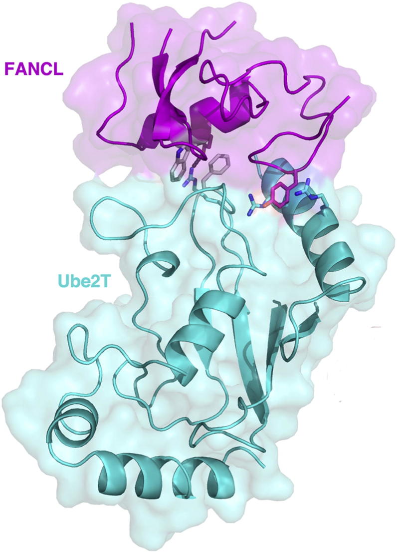
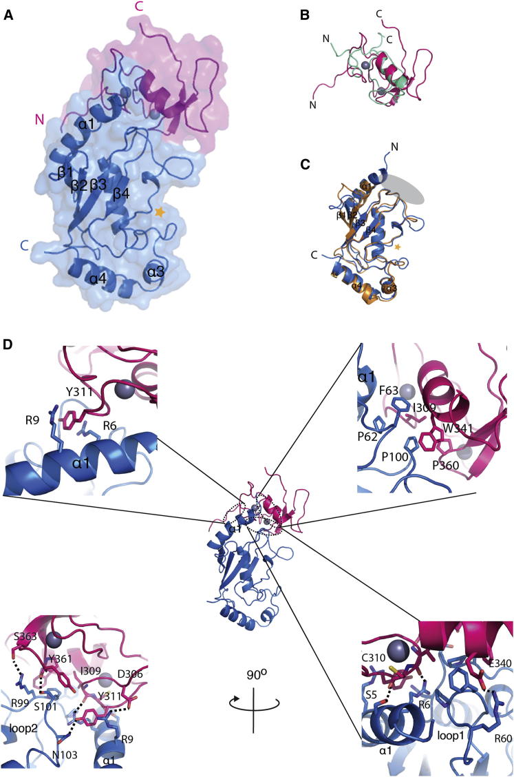
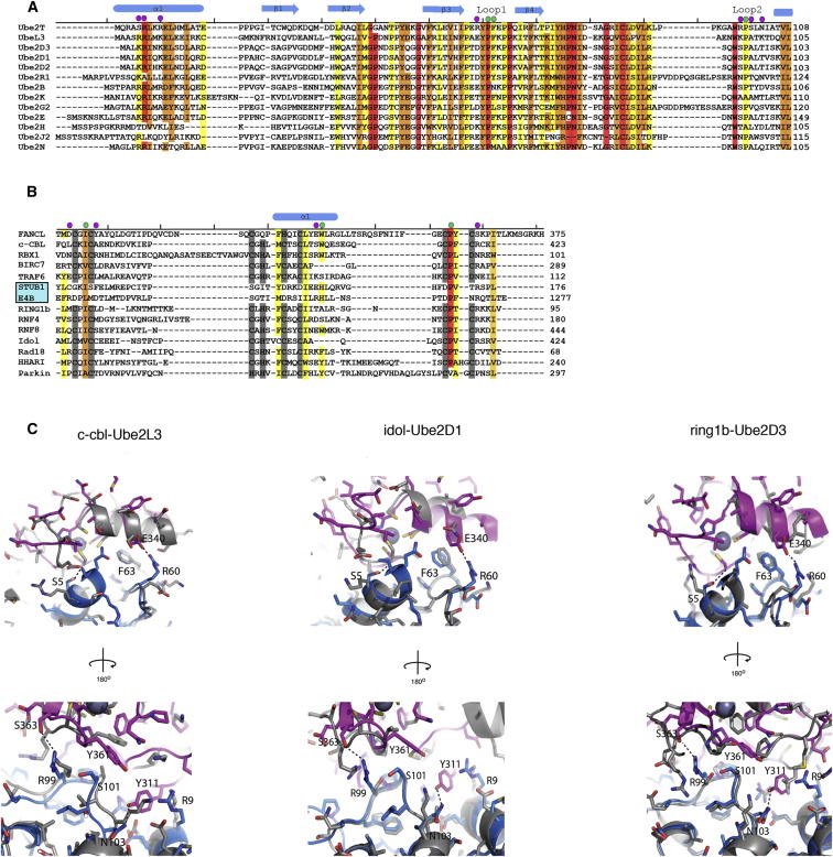
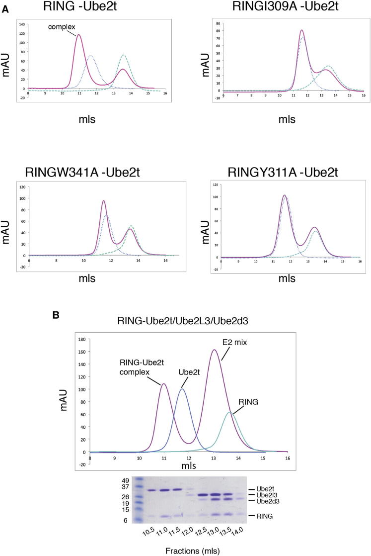
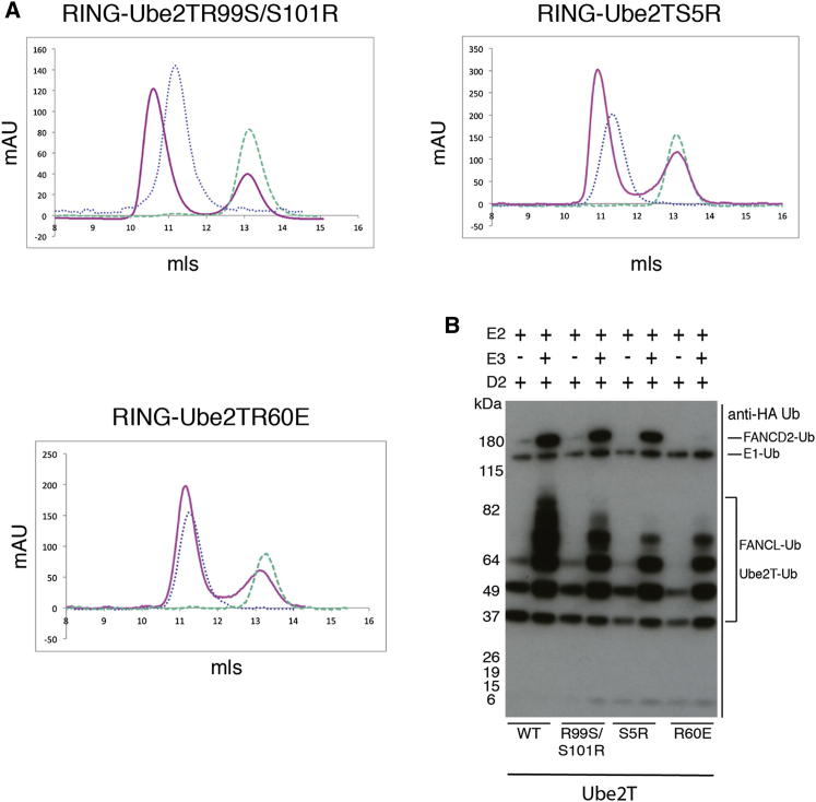
The combination of an E2 ubiquitin-conjugating enzyme with an E3 ubiquitin-ligase is essential for ubiquitin modification of a substrate. Moreover, the pairing dictates both the substrate choice and the modification type. The molecular details of generic E3-E2 interactions are well established. Nevertheless, the determinants of selective, specific E3-E2 recognition are not understood. There are ∼40 E2s and ∼600 E3s giving rise to a possible ∼24,000 E3-E2 pairs. Using the Fanconi Anemia pathway exclusive E3-E2 pair, FANCL-Ube2T, we report the atomic structure of the FANCL RING-Ube2T complex, revealing a specific and extensive network of additional electrostatic and hydrophobic interactions. Furthermore, we show that these specific interactions are required for selection of Ube2T over other E2s by FANCL.


Figures

None
Graphical abstract
Figure 1
Figure 1
Overall Structure of FANCL-Ube2T Complex (A) The overall structure of the RING domain of FANCL (magenta) bound to Ube2T (blue) is shown in cartoon representation. Gray spheres represent zinc ions. A gold star represents the position of Ube2T’s catalytic cysteine. (B) RING domain of FANCL (magenta) overlain with c-cbl RING domain (green; PDB ID code 1FBV). (C) Ube2T (blue) overlain with Ube2L3 (orange; PDB ID code 1FBV) showing the structural conservation of the UBC fold, comprising a four-stranded β-meander flanked by an N-terminal helix (helix1) and two C-terminal helices (helixes 2 and 3). A gold star represents the position of the catalytic cysteine. The gray oval shows the E3 binding interface of E2s. (D) Top left panel: The pi stacking in the binding interface between Y311 of FANCL and R6 and R9 of Ube2T. Top right panel: The hydrophobic binding interface of the RING domain (magenta) and Ube2T (blue). Bottom panels: The electrostatic and hydrogen bonding network of the RING-Ube2T interface. Interactions are represented by dashed lines. See also Figure S1.
Figure 2
Figure 2
Structural Comparison of the FANCL RING Domain-Ube2t Complex with Other RING-E2 Complexes (A) A structure-based sequence alignment of E2s. PDB ID codes of E2s, as listed in the figure: 1FBV, 3RPG, 4AP4, 4AUQ, 3RZ3, 2YB6, 3K9O, 3H8K, 2Z5D, 2F4W, 3HCT, and 3BZH. (B) A structure-based sequence alignment of RING and Ubox domains. Ubox domains are highlighted by a cyan box. PDB ID codes used of RING and Ubox domains, as listed in the figure: 1FBV, 4F52, 4AUQ, 3HCT, 2C2V, 3LIZ, 3RPG, 4AP4, 4EPO, 2YHO, 2Y43, 4KBL, and 4K7D. Residues shaded in red to yellow colors indicate conserved residues, where red corresponds to strict conservation. Gray bars indicate zinc coordinating atoms. Green circles highlight residues involved in the hydrophobic interface between FANCL and Ube2T. Purple circles denote residues involved in hydrogen bonding and electrostatic interactions in the FANCL Ube2T interface. (C) Superpositions of the FANCL RING-Ube2T complex (colored pink and blue, respectively), with c-cbl RING-Ube2L3 complex (left) shaded gray (PDB ID code 1FBV), idol-Ube2D1 complex (middle) shaded gray (PDB ID code 2YHO), and ring1b-Ube2D3 complex (right) shaded gray (PDB ID code 3RPG). Numbered residues are the same as the FANCL RING-Ube2T complex, with dashed lines showing interactions.
Figure 3
Figure 3
Conserved Hydrophobic RING Residues Are Required for Ube2T Binding and FANCL Selects Solely Ube2T In Vitro (A) Size-exclusion chromatogram profiles of wild-type (WT) or mutant RING domains (green dashed line) and WT Ube2T (blue dotted line) overlaid with profiles from binding experiments in which WT Ube2T has been incubated with WT or mutant RING domains (pink line) and subjected to size-exclusion chromatography. Binding was assessed by complex formation, which is indicated by a peak shift to the left labeled complex. (B) Size-exclusion chromatogram of FANCL RING domain incubated with an E2 mix consisting of Ube2T, Ube2D3, and Ube2L3 (pink line). Chromatograms of Ube2T (blue dotted line) and the RING domain (green dashed line) are also overlaid. A peak shift to the left is observed, indicating complex formation. SDS-PAGE gel of the fractions collected from the size-exclusion experiment and stained with Coomassie Brilliant Blue. The E2 gel bands found in the shifted peak were assessed by mass spectrometry for protein identification and confirmed as exclusively Ube2T. See also Figure S2.
Figure 4
Figure 4
Additional Residues of Ube2T Are Required for Binding the RING Domain of FANCL (A) Size-exclusion chromatogram profiles of wild-type (WT) or mutant Ube2T (blue) dotted line and WT RING domains (green dashed line) overlain with profiles from binding experiments in which WT RING domain has been incubated with WT or mutant Ube2T (pink line) and subjected to size-exclusion chromatography. Binding was assessed by complex formation indicated by a peak shift to the left. (B) An anti-HA-Ub western blot of in vitro monoubiquitination assays to assess monoubiquitination of FLAG-FANCD2 by FANCL in collaboration with different WT Ube2T and Ube2T mutants (Ube2TArg99Ser/Ser101Arg, Ube2TSer5Arg, and Ube2TArg60Glu). Lanes 2, 4, and 6 show the monoubiquitination of FLAG-FANCD2 when WT Ube2T, Ube2T Arg99Ser/Ser101Arg, and Ube2TSer5Arg are paired with FANCL. Monoubiquitination is not observed for with the Ube2TArg60Glu mutant is used (lane 8).

References

    1. Afonine P.V., Grosse-Kunstleve R.W., Adams P.D. A robust bulk-solvent correction and anisotropic scaling procedure. Acta Crystallogr. D Biol. Crystallogr. 2005;61:850–855. - PMC - PubMed
    1. Alpi A.F., Pace P.E., Babu M.M., Patel K.J. Mechanistic insight into site-restricted monoubiquitination of FANCD2 by Ube2t, FANCL, and FANCI. Mol. Cell. 2008;32:767–777. - PubMed
    1. Alter B.P. Fanconi’s anemia and malignancies. Am. J. Hematol. 1996;53:99–110. - PubMed
    1. Bailly V., Lamb J., Sung P., Prakash S., Prakash L. Specific complex formation between yeast RAD6 and RAD18 proteins: a potential mechanism for targeting RAD6 ubiquitin-conjugating activity to DNA damage sites. Genes Dev. 1994;8:811–820. - PubMed
    1. Bentley M.L., Corn J.E., Dong K.C., Phung Q., Cheung T.K., Cochran A.G. Recognition of UbcH5c and the nucleosome by the Bmi1/Ring1b ubiquitin ligase complex. EMBO J. 2011;30:3285–3297. - PMC - PubMed

Publication types

Substances