Review
. 2014 Jan 1;19(3):447-74.
doi: 10.2741/4218.

AMPK: a cellular metabolic and redox sensor. A minireview

Affiliations
Review

AMPK: a cellular metabolic and redox sensor. A minireview

Najeeb A Shirwany et al. Front Biosci (Landmark Ed). .

Abstract

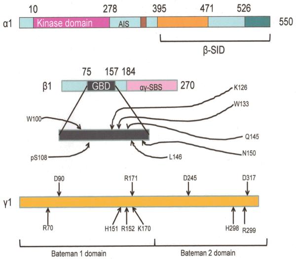
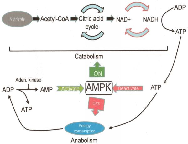
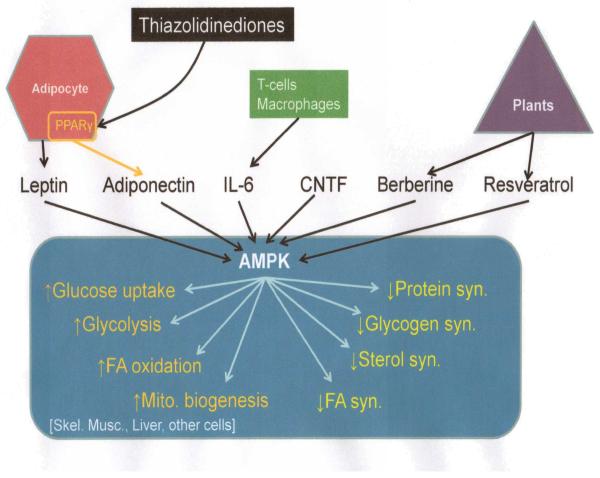
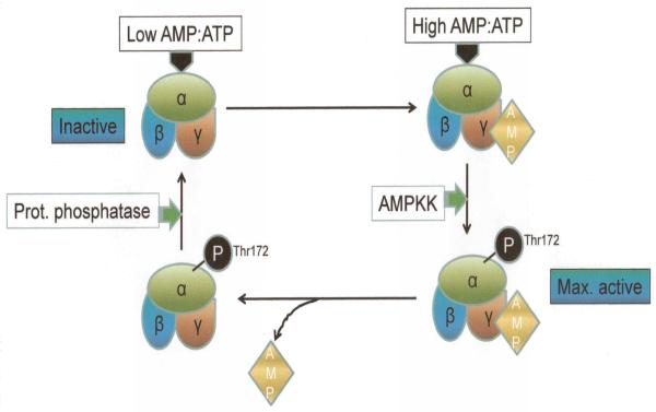
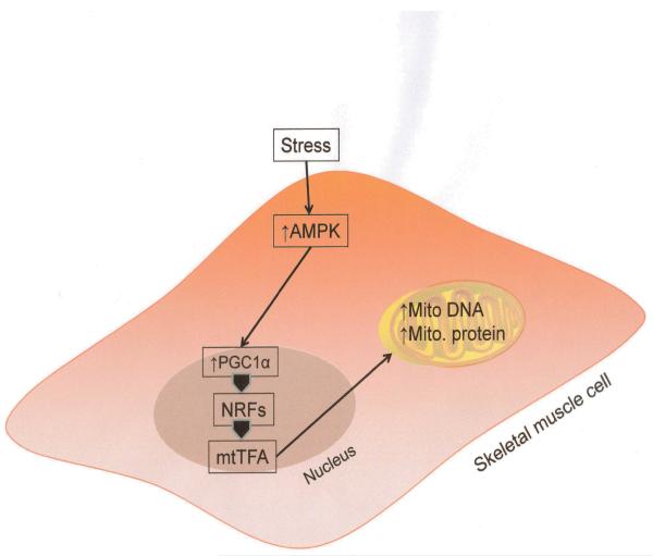
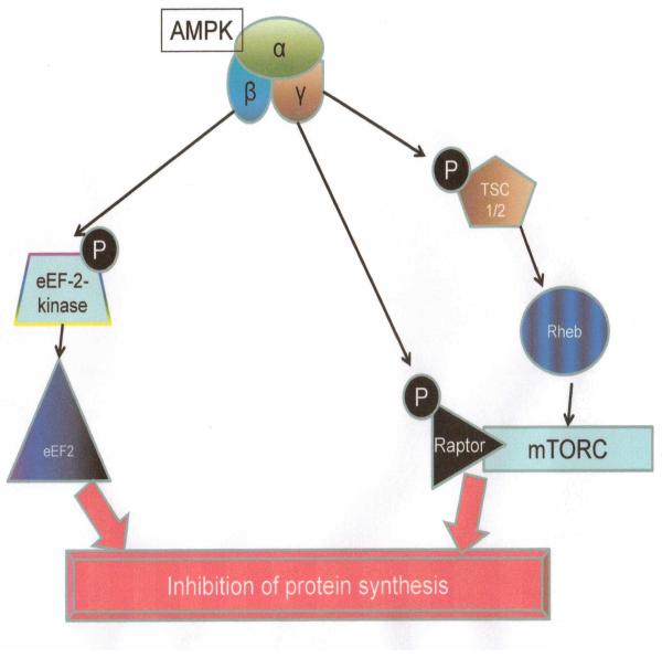
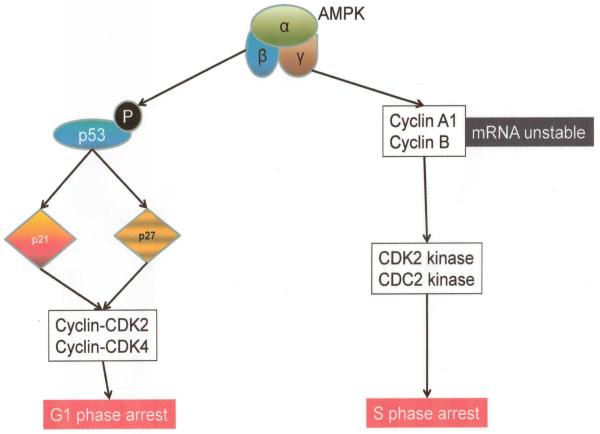
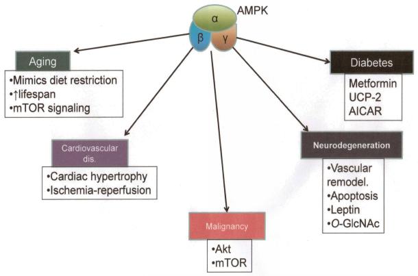
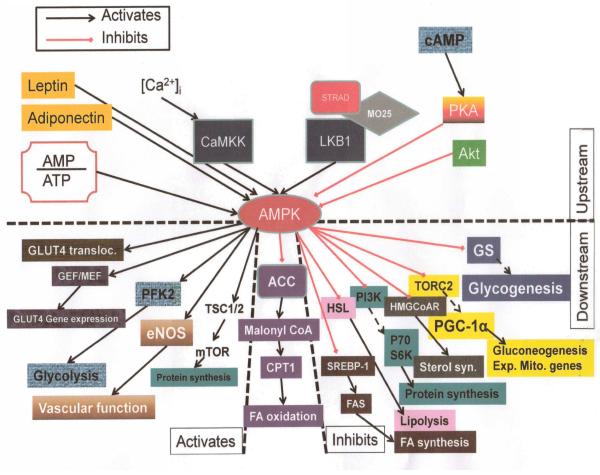
AMPK is a serine/threonine kinase that is found in all eukaryotes and is ubiquitously expressed in all organ systems. Once activated, AMPK stimulates hepatic fatty acid oxidation and ketogenesis, inhibits cholesterol synthesis, lipogenesis, and triglyceride synthesis, inhibits adipocyte lipolysis and lipogenesis, stimulates skeletal muscle fatty acid oxidation and muscle glucose uptake, and modulates insulin secretion by the pancreas. Thus its importance in many critical cellular processes is well established. For cells it is critical that energy supply and demand are closely matched. AMPK is recognized as a critical integrator of this balance. It is known to be allosterically activated by an increased AMP:ATP ratio. Activation of the kinase switches on catabolic pathways while switching off anabolic ones. It also acts as a redox sensor in endothelial cells where oxidative stress can disturb NO signaling. Abnormal NO signaling leads to disturbed vasodilatory responses. By inhibiting the formation of reactive oxygen species in the endothelium, AMPK can optimize the redox balance in the vasculature. Here, we review the role of AMPK in the cell.


Figures

Figure 1
Figure 1
Features of the AMPK subunits. Mammalian AMPK. Colored regions are ones whose structure is known. Numbers associated with α and β subunits are N- and C- terminal residues from the crystal structure. AIS: Autoinhibitory sequence. β-SID: β-subunit interacting domain. GBD: Glycogen binding domain. αγ-SBS: α and γ subunit interacting sequence. In the expanded glycogen binding domain schematic of the β subunits, letters with numbers are sites of sugar binding (pS108 is the site at Serine 108 where phosphorylation occurs). In the γ subunit, D90, R171, D245 and D317 are residues that form H-bonds with 2′ 3′-ribose hydroxyl gropus, while R70, H151, R152, K170, H298 and R299 represent basic residues that occupy the solvent accessible core of the subunit which makes contact with the nucleotide phosphates. Modified with permission from Steinberg and Kemp Physiol Rev 89:1025-1078.
Figure 2
Figure 2
Activation of AMPK by AMP:ATP ratio and influence of activated AMPK on anabolic and catabolic cellular processes.
Figure 3
Figure 3
Cellular effects of AMPK activation. A variety of triggers can activate AMPK that include adipocyte derived hormones like adiponectin, cytokines like IL-6, CNTF (ciliary neurotrophic factor) and plant derived modulators like resveratrol. Downstream effects are schematically outlined in typical target organs. Modified with permission from Hardie DG Nat Rev 2007.
Figure 4
Figure 4
Activation-deactivation cycle of AMPK. Activation of AMPK is initiated by a high AMP:ATP ratio and is triggered by AMP binding to the γ subunit and phosphorylation of Thr172 of the α subunit. This is catalyzed by AMPK kinase. The reverse (deactivation) occurs when protein phosphatase dephosphorylates the α subunit, returning AMPK to a a basal, inactive state.
Figure 5
Figure 5
AMPK activation signaling that leads to mitochondrial biogenesis. Activated AMPK signals via Peroxisome proliferator-activated receptor gamma, coactivator 1 alpha (PGC-1α), nuclear respiratory factors (NRFs) and mitochondrial transcription factors (mtTFA) to express genes encoding for mitochondrial DNA and proteins in stressed conditions. This leads to increase in mitochondrial density.
Figure 6
Figure 6
Inhibition of protein synthesis by AMPK. eEF2 kinase: Eukaryotic elongation factor 2 kinase. TSC1/2: Tuberous sclerosis complex 1-2. Rheb: Ras homolog enriched in brain. mTORC: Mammalian target of rapamycin complex.
Figure 7
Figure 7
Putative mechanism of cell cycle control by AMPK activation.
Figure 8
Figure 8
Role of AMPK in pathogenesis of various human disorders.
Figure 9
Figure 9
Integrated AMPK signaling pathways. Top half of schematic represents upstream events. Lower half represents downstream processes. In the lower half, left side are the events that are activated by AMPK while on the right are those that are inhibited by the kinase. In the center of the lower half is the main downstream effector, ACC and the consequence of its inhibition.

References

    1. Alexov EG, Gunner MR. Incorporating protein conformational flexibility into the calculation of pH-dependent protein properties. Biophys J. 1997;72(5):2075–93. - PMC - PubMed
    1. Aruksakunwong O, Wittayanarakul K, Sompornpisut P, Sanghiran V, Parasuk V, Hannongbua S. Structural and dynamical properties of different protonated states of mutant HIV-1 protease complexed with the saquinavir inhibitor studied by molecular dynamics simulations. J Mol Graph Model. 2006;25(3):324–32. - PubMed
    1. Begg DA, Rebhun LI. pH regulates the polymerization of actin in the sea urchin egg cortex. J Cell Biol. 1979;83(1):241–8. - PMC - PubMed
    1. Neuhuber WL. Lung sensors: complex functions require complex structures. Am J Respir Cell Mol Biol. 2003;28(3):265–6. - PubMed
    1. Koivusalo M, Kapus A, Grinstein S. Sensors, transducers, and effectors that regulate cell size and shape. J Biol Chem. 2009;284(11):6595–9. - PMC - PubMed

Publication types

Substances

LinkOut - more resources